❌

Reading view

Heart Health Advances with $28.7 Million Gift from Marcus Foundation

The Marcus Foundation has awarded $28.7 million to Chuck Murry, MD, PhD, director of the USC Stem Cell Center, to support a clinical trial testing a stem cell-based therapy intended to regenerate heart muscle damaged by a severe heart attack. The award, the largest the foundation has given to the University of Southern California and one of its largest contributions to academic medicine, will fund both the planned human study and a parallel research program examining how the immune system responds to transplanted cardiac cells.

The trial is scheduled to begin in 2027 at Keck Hospital of USC in partnership with the USC Cardiac and Vascular Institute. A second clinical site will be established at the University of Washington, where elements of the technology were originally developed. The first phase will enroll approximately 18 patients who have experienced extensive myocardial infarction, the medical term for a heart attack that destroys part of the heart’s muscular wall.

Patients in the study will receive stem-cell-derived heart muscle cells delivered into regions damaged by the infarction. The primary goals will be to evaluate safety and determine whether the procedure is feasible in people with severe cardiac injury. Investigators will also monitor early biological signals, including whether the transplanted cells remain active, integrate with the injured tissue and produce measurable changes in cardiac performance during recovery and long-term follow-up.

A heart attack occurs when blood flow through a coronary artery is blocked, depriving cardiac muscle of oxygen. Within minutes, cells in the affected region begin to die, and the resulting scar tissue lacks the contractile properties of healthy myocardium. Unlike tissues such as skin or blood, adult human heart muscle has only a limited capacity for self-repair. As a result, a large infarction can weaken the heart’s pumping function, increase the risk of heart failure and leave survivors unable to perform ordinary activities without fatigue or breathlessness.

Current cardiovascular treatments can reopen blocked arteries, limit further injury and help the remaining heart muscle work more efficiently. They cannot, however, routinely replace the cardiomyocytes that have already been lost. Murry’s research program is aimed at addressing that biological gap by producing replacement heart muscle cells in the laboratory and delivering them directly to damaged tissue. The strategy represents a shift from supporting a failing heart to attempting to rebuild part of its contractile apparatus.

The transplanted cells are being developed as a form of cellular therapy, in which living cells act as the therapeutic agent rather than serving merely as a vehicle for a drug. According to Murry, the cells are intended to become active within the injured heart, contribute to repair and synchronize with surrounding muscle. Achieving that outcome requires more than placing new cells into scarred tissue: the cells must survive, connect functionally with the host myocardium and avoid creating dangerous electrical disturbances or abnormal rhythms. The initial trial is therefore designed principally to establish safety before testing therapeutic effectiveness in larger patient groups.

The foundation’s funding will also support research into the immune response triggered by cell transplantation. Even cells created from stem cells can be recognized as foreign by the recipient’s immune system, potentially leading to inflammation or destruction of the transplanted population. Patients who receive cellular grafts may consequently require immunosuppressive drugs, which can increase susceptibility to infection and produce other long-term complications. Researchers will study how immune cells interact with the implanted cardiac cells and investigate methods that could reduce or eventually eliminate the need for chronic immunosuppression.

The work builds on approximately three decades of research by Murry and his collaborators to move regenerative cardiology from laboratory experiments toward clinical treatment. The program includes gene-edited cellular technology, intended to improve the compatibility or performance of therapeutic cells in the human body. The precise effectiveness of the approach will not be known until it is tested in patients, and the small 2027 cohort will not be large enough to determine whether the therapy improves survival or prevents heart failure. Those questions would require subsequent trials involving substantially more participants and longer observation.

The Marcus Foundation described the project as a potential turning point for regenerative medicine, while USC leaders said the investment could create a platform for developing additional cellular therapies. The immediate scientific challenge is to determine whether transplanted heart muscle cells can be delivered safely and function meaningfully within severely damaged human hearts. If the trial meets its safety and feasibility objectives, it could provide the clinical evidence needed to advance a new generation of treatments for myocardial infarction—therapies designed not only to help patients live with damaged cardiac tissue, but to restore some of the function that conventional medicine cannot yet replace.

Subject of Research: Stem cell-based regenerative therapy for heart muscle damaged by myocardial infarction

Article Title: $28.7 Million Award Supports USC Trial of Stem Cell Therapy to Regenerate Heart Muscle After Heart Attack

Web References: The Marcus Foundation: https://marcusfoundation.org/; USC Stem Cell Center: https://stemcell.keck.usc.edu/; Chuck Murry Lab: https://murrylab.usc.edu/charles-chuck-murry-md-phd/; USC Cardiac and Vascular Institute: https://www.keckmedicine.org/services/cardiovascular/; Keck School of Medicine of USC: https://keck.usc.edu/

Image Credits: Steve Cohn

Keywords: Heart disease, myocardial infarction, heart attack, heart muscle, stem cell research, stem cell therapy, cell therapy, regenerative medicine, cardiovascular disorders, heart failure, clinical trial, cardiac regeneration

  •  

Could Humans Regrow Limbs? Scientists Are Closer Than Ever to Unlocking a Hidden Power of Regeneration

Researchers at Texas A&M University report the successful regrowth of skeletal and connective tissues, in what they call a “critical step” toward one day enabling major tissue regeneration in humans.

Throughout time, the regenerative capabilities that, in some cases, can even restore entire lost limbs, have been viewed as an almost miraculous ability that only certain animals, such as salamanders, some reptiles, and other creatures, possess.

However, regenerative science is now unlocking the secrets of this capability, meaning that one day, even humans might be able to tap into one of nature’s most mysterious superpowers.

Awakening Humanity’s Dormant Regenerative Superpower

Current state-of-the-art research at the Texas A&M College of Veterinary Medicine and Biomedical Sciences is revealing a remarkable possibility: that regenerative abilities may not be absent in mammals after all—they may simply be dormant.

This remarkable possibility was detailed in a recent study published in Nature Communications, which details research led by Dr. Ken Muneoka with the school’s Department of Veterinary Physiology & Pharmacology (VTPP), who says he has devoted most of his career to unraveling this possibility.

Now, Muneoka and his team report a two-step treatment they developed, which successfully triggered the regeneration of bone, joint structures, ligaments, and other tissues following amputation—a significant advancement toward unlocking more powerful regenerative capabilities in humans.

“Why some animals can regenerate and others, particularly humans, can’t is a big question that has been asked since Aristotle,” Muneoka says.

Fibrolasts to the Fore

In their research, the team worked with cells that drive the healing processes and scar formation after wounds occur, which are known as fibroblasts.

In species like salamanders, similar cells go to work when injury or limb loss occurs, which begin to form into structures biologists call blastemas. These formative materials are the primary drivers behind the regrowth of tissues in such creatures.

However, unlike salamanders, mammalian cells need a bit more encouragement before they will begin to follow such regenerative pathways.

To help with this process, Muneoka and his team developed a new sequential treatment based on a pair of primary growth factors. Applying fibroblast growth factor 2 (FGF2) in an area where a wound had already healed stimulated the formation of blastema-like formations in mammal cells. Then, after several days, a bone morphogenetic protein 2 (BMP2) was applied, which similarly prompted bone cells to begin rebuilding their lost tissues.

“You first shift the cells away from scarring, and then you provide the signals that tell them what to build,” Muneoka said of this two-step process.

While the regenerated structures were not perfect replicas of the original anatomy, what Muneoka and his team achieved is still nothing short of remarkable: the team says they were able to successfully restore all major components that are typically lost when amputation occurs, including bone, tendons, ligaments, and joints.

“The Capacity is Not Absent”

Also of key significance is the fact that the team’s findings suggest that such hidden powers of regeneration might be accessible without the need for introducing external stem cells.

“You don’t have to actually get stem cells and put them back in,” Muneoka said. “They’re already there—you just need to learn how to get them to behave the way you want.”

Dr. Larry Suva, the recent study’s co-author, said the results challenge long-held assumptions about the limits of mammalian healing.

“The cells that we thought to be unprogrammable, in fact are,” Suva said. “The capacity is not absent—it’s just obscured.”

The Future of Regenerative Science

The team’s work is breaking new ground in the promising field of regenerative science, although what they have achieved so far is still in the earliest stages of what they believe may ultimately be possible.

Soon, such approaches may yield applications that could help significantly reduce scarring and may greatly help with tissue repair. Also, since BMP2 has already undergone the approval process for medical uses, and FGF2 is undergoing evaluation in clinical trials, Muneoka, Suva, and the Texas A&M team believe the path toward future medical applications may be more accessible than entirely new therapies.

“Regenerative failure in mammals can be rescued,” Muneoka said.

“Now we have a model to begin figuring out how,” he added.

The team’s recent study, “Digit regeneration in mice is stimulated by sequential treatment with FGF2 and BMP2,” was published in Nature Communications.

Micah Hanks is the Editor-in-Chief and Co-Founder of The Debrief. A longtime reporter on science, defense, and technology with a focus on space and astronomy, he can be reached at micah@thedebrief.org. Follow him on X @MicahHanks, and at micahhanks.com.

  •  

This “Zombie” Sea Creature Keeps Growing After Being Cut Apart

Man Holding Holothuria scabra Sea CucumberDetached sea cucumber tissue survived, healed, and grew for years in natural seawater, revealing a remarkable new model for regeneration and biomedical research. A chance observation has led scientists to a discovery that challenges one of biology’s most basic assumptions: that detached animal tissue eventually dies. Instead, researchers found that tissue removed from a sea [...]
  •  

Severed sea cucumber appendages don't seem to die

Organs, arms, appendages, and other complex tissues usually decay rapidly when they’re separated from their host. Over the years, biologists have seen some success with keeping them alive outside of the body—organ transplants depend on it—but it has always required germ-free environments and nutrient-rich mediums filled with growth factors. Now, though, scientists have discovered bits of tissue removed from a species of sea cucumber called Psolus fabricii can keep on living indefinitely if they’re left in ordinary seawater.

“This is naturally occurring tissue immortality,” said Sara Jobson, a researcher at Memorial University of Newfoundland and lead author of the study. “Having tissues that survive that easily is unheard of. We’ve never seen anything like this.”

The beginning of LiPfe

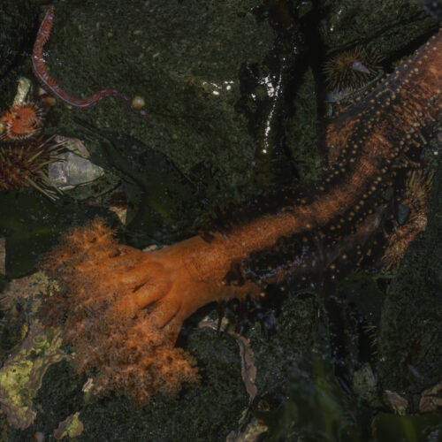
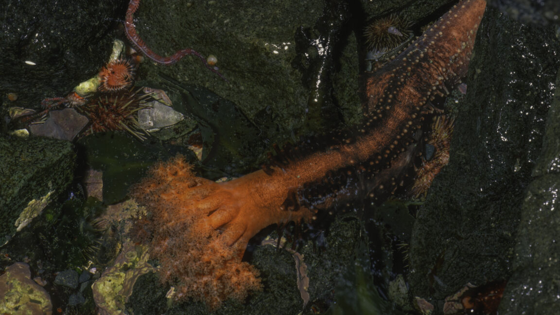
Psolus fabricii is a species of sea cucumber that lives in the cold waters of the Atlantic and Arctic oceans. Its bottom side, known as a sole, is soft and ringed by a band of tube feet that it uses to grip rocks. Once on a rock, it extends soft, branching tentacles into the water to feed on suspended particles. Because these sea cucumbers inhabit harsh environments, their feet and tentacles experience high rates of injury and loss. Evolution has therefore endowed these sites with an incredibly high capacity for regeneration.

Read full article

Comments

© Gerald Corsi

  •  
❌