❌

Reading view

Giant-nucleus cells emerge as early cancer markers in new study

For more than four decades, pathologists have glimpsed something unsettling under the microscope in the earliest stages of certain cancers: cells with nuclei far larger than they should be, swollen and distorted, lurking in tissue that otherwise appears on the verge of catastrophe. These so-called giant-nucleus cells, or karyomegalic cells, were first spotted in the 1980s in animal experiments involving iron-induced oxidative stress, yet their biological identity and their role in the genesis of cancer remained stubbornly obscure. Now, a team of researchers in Japan has provided the most detailed portrait of these cells to date, and their findings suggest that the enormous nuclei may be far more than a curiosity—they could be the founding population of early cancer, visible before any tumor exists, and potentially useful as a predictor of patient outcomes.

The study, led by Shinya Toyokuni, professor emeritus at Nagoya University Graduate School of Medicine, together with assistant professor Yingyi Kong and colleagues, was published in Redox Biology. The work addresses a deceptively simple question: when iron overload kills most cells in a tissue through oxidative damage, who survives, and what do the survivors become? Iron is an essential micronutrient, but in excess it is a potent carcinogen. Its chemistry is both the reason we need it and the reason it can harm us. Iron catalyzes the formation of hydroxyl radicals from hydrogen peroxide through the Fenton reaction, and these radicals shred DNA, oxidize lipids, and damage proteins. When the damage is severe enough, a form of programmed cell death called ferroptosis is triggered—an iron-dependent demise characterized by lipid peroxidation cascades that rupture the cell membrane. Ferroptosis has become one of the hottest topics in cancer biology precisely because it links iron metabolism, oxidative stress, and cell fate in a single mechanism.

Toyokuni’s team reasoned that if ferroptosis sweeps through iron-damaged tissue, the cells that escape it must be exceptional in some way, and those exceptions might be the seed of malignancy. To find out, they turned to a well-established animal model in which ferric nitrilotriacetate, a carcinogenic iron compound, is injected into rats, delivering a bolus of iron to the kidneys. The compound accumulates in the proximal tubules, where it catalyzes oxidative damage and, over months, produces renal cell carcinoma. In the experiments, the researchers collected kidney tissue before the injection and again one and three weeks afterward. Crucially, they also included a second experimental group: rats carrying a single mutated copy of the BRCA1 gene, the same gene that, when mutated in humans, dramatically raises the risk of breast and ovarian cancers and is associated with renal carcinoma in certain contexts.

The methodological heart of the study lies in its marriage of two technologies. The first is spatial transcriptomics, a technique that maps gene activity across individual cells within a tissue section while preserving the tissue’s original architecture—so researchers can see not only which genes are active but where in the tissue those transcriptional programs are operating. The second is computational nuclear morphometry, which quantifies the size, shape, and density of cell nuclei in the same samples. By overlaying the two datasets, the team could link the physical appearance of each cell—above all, the size of its nucleus—to its underlying transcriptional state. This integration is important because morphological atypia has long been the currency of diagnostic pathology, but connecting that atypia to molecular signatures at single-cell resolution is a recent capability.

What emerged within a week of iron administration was striking. Giant-nucleus cells appeared in the damaged kidneys, and these cells were not simply wounded cells awaiting death. They showed elevated expression of potent oncogenes, including Myc and Met, and they had acquired resistance to ferroptosis—the very cell death pathway that was eliminating their neighbors. In other words, the extreme oxidative environment had functioned as a brutal selective pressure, and the cells best adapted to survive it had activated a program that resembles, in miniature, the hallmark machinery of cancer. The enrichment of Myc, a transcription factor that drives cell growth and proliferation, and Met, a receptor tyrosine kinase involved in invasive growth, paints a picture of cells that are not merely enduring damage but reorganizing themselves around it.

The BRCA1 connection proved equally consequential. BRCA1 is best known as a guardian of the genome, a scaffold for the homologous recombination machinery that repairs double-strand DNA breaks. In the BRCA1-mutant rats, giant-nucleus cells emerged with distinctly different biological profiles from those in wild-type animals. The interpretation the researchers offer is that BRCA1 deficiency cripples DNA repair, so more of the damaged cells that would otherwise die or be repaired instead survive in their injured, genome-compromised state—becoming precancerous giant-nucleus cells. Electron microscopy and cellular respiration measurements reinforced this idea at the subcellular level: cells from BRCA1-mutant rats showed mitochondrial remodeling, with impaired iron handling and compromised respiratory function. Mitochondria, long appreciated as power plants, are also central regulators of ferroptosis and iron metabolism, and their disarray in the mutant animals may be both a consequence of BRCA1 loss and a driver of the abnormal iron biology that feeds karyomegaly.

Using their combined gene-expression and nuclear-shape data, the researchers went further and classified the giant-nucleus cells into six distinct types. One type possessed extremely large nuclei and gene signatures of stress-induced growth arrest—cells that appear to have locked themselves into a senescent-like state, consistent with profiles reported in earlier studies. Another type, found more frequently in BRCA1-mutant kidneys, showed heightened activity of cancer-related genes alongside elongated nuclei, a combination the authors interpret as a potential precancerous state. The taxonomy matters because it reframes giant-nucleus cells from a single enigmatic category into a spectrum, some members of which may be harmless or even protective while others carry genuine malignant potential.

One of the most provocative findings concerns the cells surrounding the giants. Even morphologically normal cells near giant-nucleus cells exhibited gene expression changes that appear to promote survival, suggesting that the karyomegalic cells remodel their local microenvironment in ways that extend beyond their own borders. In BRCA1-mutant kidneys, the team also documented a substantial increase in stromal cells—non-cancerous supportive cells including immune and connective tissue elements—around the damaged regions. The local tissue environment, the tumor niche in the language of modern oncology, was being reshaped long before any tumor could be called a tumor. This raises the possibility that a single conspicuous cell, easily identified by its nucleus under a conventional microscope, could serve as a sentinel for a whole neighborhood of incipient malignancy.

The leap from rat kidneys to human medicine came through two complementary analyses. First, the researchers mined TCGA-KIRC, a large public database of kidney cancer patients, asking whether tumors carrying gene-expression patterns resembling the cancer-prone giant-nucleus cells were associated with different clinical trajectories. They were: patients whose tumors shared the gene patterns of the cancer-prone giant-nucleus cells had shorter survival times, while those with patterns resembling the healthier giant-nucleus cell types had better outcomes. Not all giant-nucleus cells, in other words, are created equal, and the transcriptional fingerprints defined in the rat model appear to carry prognostic weight in human disease. Second, in a small pilot study, the team examined breast tissue from seven individuals with inherited BRCA1 mutations and fifteen without, observing abnormal nuclear features similar to those in the animal model in the mutation carriers. While the sample size is modest, the parallel hints that karyomegaly may be a cross-tissue marker of BRCA1-linked precancerous change.

The broader implications run in two directions. Clinically, the promise is that giant-nucleus cells, being easy to spot under the microscope, could function as early indicators of cancer—visible warnings in tissue that would otherwise be classified as merely damaged or inflamed. The TCGA survival data suggest that their gene-expression signatures might even stratify patients by risk once a tumor has formed. Scientifically, the study offers a concrete mechanism connecting an environmental exposome factor—iron-catalyzed oxidative stress—to a defined, transcriptionally characterized precancerous population, with ferroptosis resistance as the pivot. It also implies testable therapeutic hypotheses: if blocking mitochondrial remodeling or reversing ferroptosis resistance could prevent the persistence of the precancerous cells, early intervention might stop cancer before it starts. Toyokuni and his colleagues are careful to note that long-term studies will be needed to determine how giant-nucleus cells actually progress into tumor cells, whether such interventions can work, and whether nuclear shape combined with gene patterns can be validated as a diagnostic in larger patient cohorts. But the central message is already legible: some of the earliest chapters of cancer may be written in cells that have grown too large to be ignored.

Subject of Research: Animals

Article Title: Iron-catalyzed oxidative stress reveals an exposome-related ferroptosis-resistant karyomegalic niche in BRCA1-linked renal carcinogenesis.

Article References: Kong, Y., Shiraki, Y., Furuhashi, K., Maruyama, S., Imaoka, T., Enomoto, A., & Toyokuni, S. (2026). Iron-catalyzed oxidative stress reveals an exposome-related ferroptosis-resistant karyomegalic niche in BRCA1-linked renal carcinogenesis. Redox Biology, 95, Article 104293. https://doi.org/10.1016/j.redox.2026.104293

Image Credits: AI Generated

DOI: 10.1016/j.redox.2026.104293

Keywords: giant-nucleus cells, karyomegaly, ferroptosis, BRCA1, iron-catalyzed oxidative stress, spatial transcriptomics, renal carcinogenesis, mitochondrial remodeling, nuclear morphometry, precancerous cells, TCGA-KIRC, Nagoya University

  •  

244-Million-Year-Old Fossil Preserves Earliest Reptilian Digestive System

Life reconstruction of Austronaga minuta. Image credit: Gabriel Ugueto.

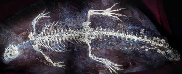
An exquisitely preserved specimen of Austronaga minuta, a small, long-necked marine reptile from the Triassic of China, contains a fossilized stomach, liver and intestines, offering paleontologists their oldest detailed glimpse yet of how early reptiles digested food.

The post 244-Million-Year-Old Fossil Preserves Earliest Reptilian Digestive System appeared first on Sci.News: Breaking Science News.

  •  

Brain-region microstructure and gene activity reveal Alzheimer’s neurodegeneration

Alzheimer’s disease may be leaving a more precise anatomical signature in the brain than scientists once realized. A new study published in Translational Psychiatry investigates regional changes in the fine structure of brain tissue immediately beneath the cerebral cortex, known as the juxtacortical region, and links those changes to patterns of gene expression associated with neurodegeneration. The work, led by J. Zhang, L. Sun, X. Yang and colleagues, offers a combined view of Alzheimer’s biology that connects what can be detected through brain imaging with the molecular programs active inside vulnerable neural tissue.

The juxtacortical zone is a narrow but biologically important interface. It lies directly beneath the cortex, the outer layer responsible for functions including memory, language, perception and decision-making. Although much Alzheimer’s research has focused on cortical thinning, large-scale tissue loss and abnormalities in deeper brain structures, the tissue bordering the cortex may reveal earlier or more regionally specific changes. Its organization includes densely packed axons, dendrites, neurons, glial cells and blood vessels, all of which can be affected as the disease progresses. Subtle disruption in this area could therefore provide clues before dramatic anatomical damage becomes visible.

The study’s central idea is that Alzheimer’s disease does not alter every brain region in the same way. Instead, neurodegeneration follows a complex geography shaped by cellular composition, connectivity, metabolism and genetic vulnerability. By examining regional juxtacortical microstructural changes, the researchers seek to move beyond a simple question of whether tissue has shrunk. Microstructure refers to the organization of brain components at scales smaller than conventional anatomical imaging can directly resolve. Variations in water diffusion, tissue organization and local architecture can serve as indirect markers of changes to axons, membranes, extracellular space and other microscopic features.

This regional perspective matters because the same imaging signal can arise from different biological processes. A change in tissue organization may reflect loss of neuronal connections, damage to myelinated fibers, altered glial activity, inflammation or shifts in the balance of cellular compartments. Imaging alone can reveal where abnormalities are concentrated, but it cannot always explain why they occur. The researchers therefore paired their investigation of juxtacortical structure with gene-expression information, creating a bridge between regional anatomy and molecular biology. That combination can help identify which biological pathways may underlie the imaging patterns seen in Alzheimer’s disease.

Gene expression describes the activity of genes within cells and tissues: which genetic instructions are being read, and at what levels. In the Alzheimer’s brain, these molecular signatures can point toward processes such as synaptic dysfunction, immune activation, impaired energy production, protein handling and cellular stress. Mapping gene-expression patterns onto regions showing microstructural alterations may reveal whether vulnerable areas share particular molecular characteristics. It may also help distinguish tissue changes related directly to neurodegeneration from those associated with aging or general brain atrophy. The result is a more layered portrait of disease, linking microscopic organization to the biological machinery that maintains it.

The findings described by the paper’s title suggest that these two forms of evidence—regional microstructure and gene activity—provide complementary insights into Alzheimer’s pathology. Rather than treating the brain as a uniform organ undergoing gradual damage, the research emphasizes a mosaic of vulnerability. Some areas may show distinctive structural disruption alongside gene-expression profiles associated with neuronal injury or altered support-cell function, while other regions may be relatively resilient. Such differences could help explain why symptoms vary between individuals and why memory, language, attention and other abilities can deteriorate along different trajectories.

The work could also influence how scientists think about biomarkers. A biomarker is a measurable biological feature that helps indicate disease presence, progression or response to treatment. Traditional markers of Alzheimer’s include abnormal amyloid and tau proteins, brain volume loss and changes in metabolism. Juxtacortical microstructural measures, especially when interpreted alongside molecular data, could eventually add regional detail to this toolkit. They might help researchers identify tissue at risk, track the spread of degeneration or evaluate whether an experimental therapy is protecting neural architecture even before clinical improvement becomes obvious.

However, the study should not be interpreted as proof that a single imaging signature can diagnose Alzheimer’s disease or predict an individual’s future with certainty. Microstructural measurements are indirect, and gene-expression data often come from specialized datasets that may not capture every cell type or every stage of disease. Differences in age, disease severity, medication, vascular health and individual brain organization can also influence results. The strength of the approach lies not in one isolated measurement, but in the convergence of regional imaging and molecular evidence. Together, these methods can generate testable hypotheses about how and where neurodegeneration unfolds.

The broader message is that Alzheimer’s research is moving toward increasingly integrated maps of the human brain. The study by Zhang, Sun, Yang and colleagues presents the juxtacortical region as a potentially informative frontier, where microscopic tissue organization and gene regulation intersect. If future research confirms these relationships in larger and more diverse groups, the approach could sharpen disease staging and improve the design of targeted therapies. For now, the findings add weight to a crucial principle in neuroscience: understanding Alzheimer’s requires knowing not only which brain regions are damaged, but also how their cellular architecture and molecular identity make them vulnerable.

Subject of Research: Regional juxtacortical microstructural changes and gene expression in Alzheimer’s disease

Article Title: Regional juxtacortical microstructural changes and gene expression provide insights into neurodegeneration in Alzheimer’s disease

Article References: Zhang, J., Sun, L., Yang, X. et al. “Regional juxtacortical microstructural changes and gene expression provide insights into neurodegeneration in Alzheimer’s disease.” Translational Psychiatry (2026). https://doi.org/10.1038/s41398-026-04292-z

Image Credits: AI Generated

DOI: https://doi.org/10.1038/s41398-026-04292-z

Keywords: Alzheimer’s disease, neurodegeneration, juxtacortical microstructure, gene expression, brain imaging, neuroscience, biomarkers, translational psychiatry

  •  

Brown fat microRNAs mapping shows secreted signaling network between organs

A newly published study in Nature Metabolism reports that brown fat can act as a hub of molecular messaging, using secreted microRNAs to coordinate physiology across the body. Researchers focused on brown adipose tissue—well known for its role in thermogenesis—and asked whether it also participates in long-distance communication via RNA signals.

To visualize and track these messages, the team employed brown-fat–specific RNA labeling in living systems. This strategy allowed them to connect RNA production within adipocytes to downstream effects elsewhere, moving beyond correlations toward a clearer map of inter-organ RNA traffic.

The central finding is the presence of a communication network mediated by microRNAs released from brown fat. These small regulatory RNAs appear to travel beyond the tissue of origin and engage gene-regulatory pathways in other organs, effectively relaying information about metabolic state.

Functionally, the work supports a model in which microRNA “dispatch” from thermogenic fat tunes cellular programs in remote targets. Such programs likely involve post-transcriptional regulation, a process microRNAs are known to influence by pairing with complementary sequences in messenger RNAs.

Importantly, the approach does not treat microRNAs as generic circulating molecules. Instead, the labeling confirms their brown-fat origin and strengthens the link between secretory RNA dynamics and systemic physiological outcomes.

The study also highlights the specificity of the messaging: not all RNAs are equally represented, and the labeled microRNA repertoire points to organized, tissue-defined signaling rather than random leakage.

Beyond mapping pathways, the results carry implications for metabolic disease research. If brown fat microRNAs can reshape gene expression in other organs, manipulating these signals could offer a route to modulate energy balance, insulin sensitivity, or inflammatory tone.

For now, the findings provide a blueprint for how RNA labelling combined with tissue specificity can uncover hidden layers of endocrine-like communication. As techniques improve, the field may move toward a broader “RNA connectome” linking organ behavior through secreted regulatory molecules.

Researchers describe their results as evidence that brown fat is not only a metabolic engine but also an information broadcaster. In the evolving landscape of viral-style science news, this discovery underscores how tiny RNA molecules can orchestrate large-scale biological conversations.

Subject of Research: Inter-organ communication via secreted microRNAs from brown adipose tissue.

Article Title: Brown fat-specific RNA labelling reveals a network of inter-organ communication by secreted microRNAs.

Article References: Lino, M., Palermo-Ruiz, G., Efthymiou, V. et al. Brown fat-specific RNA labelling reveals a network of inter-organ communication by secreted microRNAs. Nat Metab (2026). https://doi.org/10.1038/s42255-026-01558-0

Image Credits: AI Generated

DOI: https://doi.org/10.1038/s42255-026-01558-0

  •  

113-Million-Year-Old Pterosaur Fossil Reveals What Flying Reptiles Ate

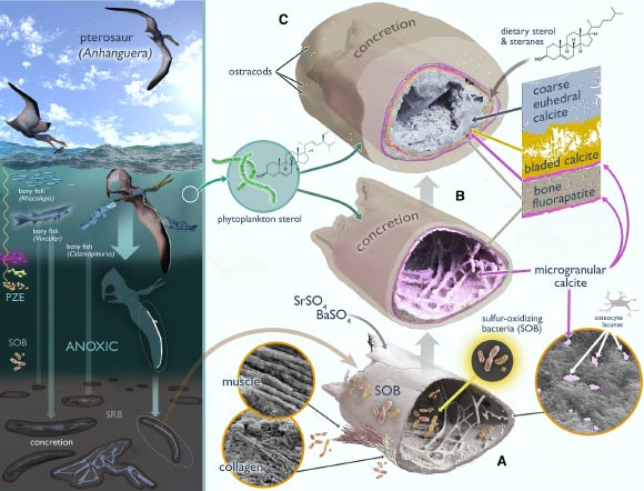
Grice et al. integrate organic geochemical analyses with high-resolution micro-mineral imaging of a three-dimensionally preserved Cretaceous pterosaur wing phalanx from Brazil to reveal steroid biomarkers and multi-stage mineralization pathways underlying its preservation. Image credit: Grice et al., doi: 10.1016/j.isci.2026.116199.

A 113-million-year-old pterosaur fossil from northeastern Brazil has yielded rare evidence of soft tissues, organic molecules and chemical traces of a diet heavy in fish and cephalopods such as squid or nautilus relatives.

The post 113-Million-Year-Old Pterosaur Fossil Reveals What Flying Reptiles Ate appeared first on Sci.News: Breaking Science News.

  •  

125-Million-Year-Old Crocodile Relative Reveals Its True Colors

The holotype specimen of Montsecosuchus depereti. Image credit: Castillo-Visa et al., doi: 10.1093/zoolinnean/zlag076.

Exceptionally preserved skin of Montsecosuchus depereti, an extinct crocodylomorph no larger than a house cat that prowled the tropical wetlands of Early Cretaceous Spain, has allowed paleontologists to reconstruct details of its scales, sensory organs and even possible banded markings along its tail.

The post 125-Million-Year-Old Crocodile Relative Reveals Its True Colors appeared first on Sci.News: Breaking Science News.

  •  

New Study Reveals Ebola Virus Persistence in the Central Nervous System, Led by Mount Sinai Microbiologists

Ebola Virus Finds Sanctuary in the Brain: Breaking Ground with Human Cerebral Organoids

In a landmark study published in Nature Microbiology, researchers from the Icahn School of Medicine at Mount Sinai and the Bernhard Nocht Institute for Tropical Medicine (BNITM) have uncovered how the deadly Ebola virus can persist and replicate stealthily within the human central nervous system. By leveraging cutting-edge cerebral organoid models—three-dimensional brain-like structures derived from human pluripotent stem cells—the team has shed light on the viral mechanisms that allow long-term survival, raising critical implications for treatment and prevention strategies of Ebola virus disease (EVD).

It has long been a harrowing clinical observation that survivors of acute Ebola infections sometimes suffer relapses or develop inflammatory syndromes months or even years after recovery. This enduring threat is attributed to the virus’s ability to hide out in “immune-privileged” regions of the body—such as the central nervous system (CNS)—where the immune surveillance is deliberately restrained to protect vulnerable tissue from damaging inflammation. This latest research elucidates the ways Ebola virus exploits this sanctuary and endures prison-like conditions inside cerebral tissue without fully succumbing to immune clearance.

The team engineered cerebral organoids by coaxing human induced pluripotent stem cells (iPSCs) into forming spherical, differentiated structures composed of multiple neural cell types: neurons, astrocytes, and microglia alike. These organoids mimic the cellular environment of the human brain closely enough to serve as a viable model to explore viral persistence with unprecedented resolution and relevance. Unlike traditional animal models, these human-derived organoids enable direct examination of host-virus interactions in a context that recapitulates human neuroanatomy and immunology.

Longitudinal infection experiments revealed that Ebola virus and related filoviruses—including Sudan, Reston, and Marburg viruses—could sustain active replication within these cerebral organoids for up to 120 days. More intriguingly, Ebola virus infected not just neurons but also astrocytes and microglia, the brain’s resident immune cells. The virus exhibited two spreading modalities: direct cell-to-cell contact transmission as well as classical budding from host cells. This productive persistence clearly places the virus in an infectious, replicative state rather than a latent or inactive one.

Despite the robust production of pro-inflammatory cytokines by infected organoids, the innate immune responses orchestrated within this model failed to clear the infection entirely. The study observed an upregulation of immune and inflammatory markers at later stages, mirroring clinical presentations seen in Ebola virus disease survivors who develop delayed meningoencephalitis and other inflammatory sequelae. The data affirm that persistent filovirus infection in immune-privileged tissues contributes to localized inflammation, fostering neurological disease conditions even after primary viremia subsides.

At the molecular level, whole-genome sequencing of viral populations recovered from persistently infected organoids uncovered the presence of defective viral genomes (DVGs) and a spectrum of mutations emerging during chronic replication. DVGs are truncated or mutated forms of viral genomes known to modulate replication dynamics and immune evasion in other viral families. These defective genomes may allow the Ebola virus to dampen replication while maintaining a foothold inside host tissue, facilitating long-term survival without triggering overwhelming immune destruction.

Several mutations identified in the viral sequences corresponded to variants previously reported to attenuate replication in natural infections, while others were novel and uncharacterized. This emerging mutational landscape hints at an ongoing evolutionary arms race as the virus adapts to the CNS environment and immune pressures. Such adaptations could be critical determinants in the capacity of Ebola virus to establish persistent reservoirs and cause relapses.

This cerebral organoid model’s faithful recapitulation of human Ebola virus infection patterns underscores its utility as a powerful platform for mechanistic studies of viral persistence. Unlike animal models, which often fail to mimic intricate human neural immunology adequately, organoids offer a nearer-to-human experimental system. This breakthrough has potential ramifications for testing and optimizing targeted antiviral therapies aimed specifically at reservoirs residing in immune-privileged compartments.

Moreover, cerebral organoids provide an ethically favorable and scalable alternative to animal experiments, fostering more humane and potentially faster progress in infectious disease research. This innovation aligns with global efforts to reduce animal use in biomedical studies and to increase translational fidelity to human conditions.

Looking ahead, the research team emphasizes expanding their investigations to include other lesser-studied filoviruses such as Taï Forest, Bombali, and Bundibugyo viruses. Comprehensive deciphering of host–virus dynamics in such a model may unravel universal persistence mechanisms across filovirus species, informing cross-cutting therapeutic strategies. Crucially, this work also lays groundwork for exploring how viral persistence in the CNS intersects with immune modulation, neuroinflammation, and long-term neurological sequelae in EVD survivors.

Persistent Ebola infection in the brain highlights a formidable challenge for public health, given the potential for virus reactivation or even transmission from immune-privileged sanctuaries. This study delivers a compelling window into that hidden battleground, leveraging state-of-the-art human cellular models to confront a deadly pathogen’s stealth strategies. Unlocking the biology of Ebola’s cerebral persistence may ultimately pave the way for interventions that not only save lives during acute infection but also circumvent post-recovery complications and community outbreaks.

As our understanding deepens, harnessing cerebral organoids as investigative tools promises to revolutionize the study of neurotropic viral infections. The Mount Sinai and BNITM collaboration exemplifies how multidisciplinary, translational research can illuminate the dark corners of viral pathogenesis and guide clinical innovation for diseases once deemed intractable.


Subject of Research: Animals (via human-derived cerebral organoids)

Article Title: Host–virus determinants of Ebola virus persistence in a human cerebral organoid model

News Publication Date: 12-Jun-2026

Web References:
https://www.nature.com/articles/s41564-026-02388-2
http://dx.doi.org/10.1038/s41564-026-02388-2

Keywords: Ebola virus, cerebral organoids, central nervous system, viral persistence, filoviruses, neuroinflammation, antiviral therapy, immune evasion, defective viral genomes, virus-host interaction

  •  

This “Zombie” Sea Creature Keeps Growing After Being Cut Apart

Man Holding Holothuria scabra Sea CucumberDetached sea cucumber tissue survived, healed, and grew for years in natural seawater, revealing a remarkable new model for regeneration and biomedical research. A chance observation has led scientists to a discovery that challenges one of biology’s most basic assumptions: that detached animal tissue eventually dies. Instead, researchers found that tissue removed from a sea [...]
  •  
❌