❌

Reading view

iPhone 18 Pro adds variable aperture, A20 Pro, and longer battery life

iPhone 18 Pro arrives with Apple concentrating its biggest changes in three areas: camera control, sustained performance, and battery life. The new Pro models retain familiar 6.3-inch and 6.9-inch display sizes, but introduce a variable-aperture main camera, the 2-nanometer A20 Pro chip, redesigned thermal hardware, and a smaller Dynamic Island.

The lineup consists of iPhone 18 Pro and iPhone 18 Pro Max, starting at $1,199 and $1,299 in the U.S., respectively. Both begin with 256GB of storage and are available in black, silver, glacier, and a new burgundy finish. Preorders opened September 12, with retail availability beginning September 18 in more than 65 countries and regions.

Rather than treating the Pro models as a cosmetic update, Apple has focused on changes that become more noticeable during demanding use, particularly photography, video work, gaming, AI processing, and long sessions away from a charger.

iPhone 18 Pro brings variable aperture to the main camera

The centerpiece is a new 48MP Fusion Main camera with a physical variable aperture, a first for the iPhone.

Six laser-cut aperture blades can adjust the amount of light reaching the sensor while also changing depth of field. The camera supports four aperture settings: ƒ/1.48, ƒ/1.8, ƒ/2.8, and ƒ/4.0.

At the wider end, the camera can gather more light in darker scenes. A narrower aperture can keep more people or background detail in focus, giving photographers another tool beyond computational depth effects and Portrait mode.

Apple also gives users direct access to those settings through new Pro controls in the Camera app. Aperture, shutter speed, and white balance can be adjusted manually, while a histogram provides a more conventional way to monitor exposure.

The feature is paired with a new main-camera sensor and updated computational imaging pipeline. Apple says the combination improves low-light performance and detail, while Photographic Styles gain texture and grain controls for additional control over an image’s appearance.

Video receives several additions as well. Cinematic effects can now be applied after recording to footage captured at up to 60 frames per second, while Time-lapse can record in 4K with Dolby Vision HDR. Audio Mix has also been updated with improved voice processing and the ability to isolate music.

For photographers who want more evidence of how an image originated, Apple is introducing Apple Reference Image. In Reference mode, the main camera signs sensor data, which Private Cloud Compute uses to create an unalterable reference version that can be compared with an edited image in Photos. Apple is also preparing support for the SynthID standard as another way of identifying AI-generated or altered content.

Close-up of a camera lens with visible aperture blades, surrounded by a maroon-colored body—reminiscent of the sophisticated design seen on the iPhone 18 Pro Max. Subtle reflections are visible on the lens surface.
Image Credit: Apple Inc.

iPhone 18 Pro uses A20 Pro and a redesigned vapor chamber

A20 Pro is built using a 2-nanometer manufacturing process and contains a six-core CPU, seven-core GPU, and dual 16-core Neural Engine.

Apple says memory bandwidth is 50% higher than on A19 Pro, while GPU performance can be up to 40% faster. The Neural Engine provides 32 cores in total, doubling the AI processing capability of the previous Pro chip.

The silicon change is closely tied to a redesigned cooling system. Apple has rearranged the chip package so that the silicon die sits beside the memory rather than beneath it in the thermal path, allowing A20 Pro to connect more directly to a next-generation vapor chamber.

That vapor chamber has three times the surface area of the previous generation and uses new materials to move heat away from the processor more effectively. Apple claims the design can deliver up to a 40% gain in sustained performance compared with iPhone 17 Pro.

That distinction is more relevant than peak benchmark numbers for workloads such as extended gaming sessions, video editing, computational photography, and local AI models, where heat can force a phone to reduce performance over time.

Connectivity has also moved further toward Apple-designed silicon. The N1 wireless chip supports Wi-Fi 7, Bluetooth 6, and Thread, while the new C2 modem succeeds C1X. Apple says C2 uses 15% less energy while offering faster uploads and adds mmWave support in the U.S.

A metallic chip with the Apple logo and text “A20 PRO” in bold, centered on a dark background with a subtle multicolored glow around the edges—designed to power the next-generation iPhone 18 Pro and iPhone 18 Pro Max.
Image Credit: Apple Inc.

iPhone 18 Pro Max reaches 45 hours of video playback

Battery improvements are another major part of the new hardware.

Apple rates eSIM-only versions of iPhone 18 Pro for up to 36 hours of video playback, while iPhone 18 Pro Max reaches as much as 45 hours. Apple says Pro Max receives the largest battery-life increase ever delivered in an iPhone.

Some of that gain comes from using the space previously occupied by a physical SIM tray. In markets receiving eSIM-only models, including the U.S., Canada, Japan, Mexico, Saudi Arabia, and the UAE, the larger internal battery provides an additional two hours of rated video playback.

Charging has accelerated as well. iPhone 18 Pro can reach 50% charge in approximately 15 minutes using a supported wired setup, while wireless charging reaches 50% in around 30 minutes. Apple says five minutes of wired charging on iPhone 18 Pro Max can provide roughly seven hours of video playback.

Both devices use aluminum unibody construction with Ceramic Shield 2 on the front and Ceramic Shield on the back. Storage options run from 256GB through 2TB.

The iPhone 18 Pro weighs 211 grams, while iPhone 18 Pro Max weighs 249 grams. Display sizes remain 6.3 inches and 6.9 inches, respectively.

Four iPhones—featuring the iPhone 18 Pro and iPhone 18 Pro Max—in black, white, blue, and deep red colors are displayed upright in a row against a black background, showing their backs and triple camera arrays.
Image Credit: Apple Inc.

iOS 27 brings Siri AI and new Apple Intelligence tools

The hardware launches alongside iOS 27, which places Apple Intelligence and the new Siri AI more prominently across the system.

Siri AI can use personal context from information stored across apps, respond to onscreen content, perform actions in supported apps, and handle more open-ended questions. A new Siri mode in Camera lets users ask questions and take action based on objects or information visible through the viewfinder.

Photo tools include Spatial Reframing, Extend, and an upgraded Clean Up feature, while Safari can monitor selected webpages for changes such as a product restock or price adjustment.

The Dynamic Island has also been physically reduced and redesigned. It can display up to three Live Activities simultaneously while continuing to support Face ID.

Apple released iOS 27 on September 14, while Siri AI is rolling out initially as a beta in English, with additional language support planned. iPhone 18 Pro and iPhone 18 Pro Max reach stores September 18, giving Apple a Pro lineup whose most substantial additions are less about a new exterior shape and more about physical camera control, heat management, and how long the hardware can sustain demanding workloads.

The post iPhone 18 Pro adds variable aperture, A20 Pro, and longer battery life appeared first on AppleMagazine.

  •  

Spanish Amber Deposit Offers Rare Window into Cretaceous Life

Life reconstruction of Hallucinochrysa diogenesi. Image credit: J.A. Peñas.

El Soplao, a fossil site tucked into the hills of Cantabria in northern Spain, stands out for yielding amber pieces with a blue-violet hue and abundant bioinclusions, according to a new review paper.

The post Spanish Amber Deposit Offers Rare Window into Cretaceous Life appeared first on Sci.News: Breaking Science News.

  •  

Simulating Fluid–Structure Interaction in Eyes with Detached Descemet Membrane

In a finding that could reshape how ophthalmologists think about one of the most delicate complications of corneal surgery, a team of computational engineers has built the first detailed computer model of a human eye in which the Descemet membrane—the paper-thin basement layer at the back of the cornea—has become detached. By simulating the fluid–structure interaction between the aqueous humor sloshing inside the eye’s anterior chamber and the flexible, detached membrane itself, the researchers have produced quantitative predictions about the mechanical stresses such a detachment imposes, and about how different transplanted corneas might alter those stresses during post-operative thermal therapy.

The study, published in the Annals of Biomedical Engineering, addresses a long-standing blind spot in ocular biomechanics. Detachment of the Descemet membrane is a recognized complication of cataract surgery, corneal transplantation and certain corneal dystrophies, and it can cause significant corneal swelling and vision loss if the membrane does not reattach. Yet most previous computational examinations of the anterior chamber have treated the eye’s interior as rigid-walled plumbing, focusing on fluid flow or heat transfer while excluding the complex two-way coupling between the intraocular fluid and the fragile ocular structures suspended within it. The new work changes that by modeling the detached Descemet membrane as a deformable, elastic solid that bends, stretches and deforms under the pressure and shear of the aqueous humor flowing around it.

The researchers, led by Ammar I. Alsabery of the Islamic University in Najaf, Iraq, together with colleagues from the University of Basrah, King Khalid University in Saudi Arabia, Saveetha School of Engineering in India, and Universiti Kebangsaan Malaysia, employed a coupled numerical scheme that solves simultaneously for the buoyancy-driven flow of aqueous humor and for the structural deformation of the membrane. The geometry represents the anterior chamber—the fluid-filled space between the cornea and the iris—with a warm region corresponding to the lens contact zone held at an isothermal hot temperature, mimicking the thermal gradients that arise in ocular warming therapies and in normal physiological conditions.

At the heart of the analysis is the Rayleigh number, a dimensionless parameter that governs the strength of natural convection: warmer, less dense fluid rises while cooler fluid sinks, generating circulating currents within the chamber. The team varied the Rayleigh number across three orders of magnitude, from 10³ to 10⁶, which corresponds to temperature differences spanning from a fraction of a degree to 250 Kelvin. This range deliberately encompasses both the mild thermal gradients present in the living eye and the much more aggressive temperature differences applied during clinical thermal therapy of the cornea, allowing the same model to speak to both resting physiology and active treatment.

Equally central to the study is the elasticity modulus of the detached membrane. The researchers swept this property across nine orders of magnitude, from 10⁸ to 10¹¹, a range designed to represent everything from highly compliant, freshly detached tissue to the much stiffer mechanical character of donor corneal tissue used in transplantation. Because the Descemet membrane’s stiffness varies with age, disease state and surgical history, this parametric approach lets the model answer a question that clinicians genuinely care about: does the type of cornea grafted into a patient change the mechanical load experienced by a detached membrane during recovery? The answer, according to the simulations, is nuanced. The cornea type does affect the stress imparted on the detached Descemet membrane, but it does not affect the thermal characteristics of the anterior chamber or the overall displacement behavior within it. In other words, stiffer transplanted tissue changes the mechanical environment at the membrane itself without appreciably altering the convective heat transfer patterns of the aqueous humor circulating beneath it.

The fluid dynamics of the anterior chamber turned out to be far more sensitive to geometry than to tissue stiffness. One of the most striking results concerns the iris, the pigmented diaphragm whose length in the model determines the extent of the hot, isothermal contact region associated with the lens. When the team simulated a shrinkage of the iris—effectively reducing the length of the contacting part of the lens held at the hot temperature—they found that the convective thermal characteristic of the anterior chamber rose by approximately 78 percent. This dramatic enhancement arises because the reduced iris length reshapes the buoyancy-driven circulation cells, allowing warmer fluid to travel more freely and exchange heat more efficiently with its surroundings. For clinicians, the implication is that the geometry of the anterior chamber, shaped in part by the iris and the lens, is a first-order determinant of how heat is distributed during thermal therapy, potentially more important than the material properties of the graft itself.

The temperature difference applied during thermal therapy also proved to be a powerful lever. Raising the applied thermal gradient from 2.5 Kelvin to 25 Kelvin—a tenfold increase—promoted heat transfer within the anterior chamber by 47.6 percent. While the scaling is sublinear rather than directly proportional to the temperature difference, the result confirms that modest increases in therapeutic temperature can yield meaningful gains in convective heat delivery without requiring a hundredfold jump in applied thermal load. The finding offers a quantitative framework for calibrating thermal treatments of the anterior segment: therapy protocols could, in principle, be tuned to achieve target heat transfer rates while respecting the narrow thermal safety margins of living ocular tissue.

Technically, the simulations belong to a class of problems known as fluid–structure interaction, or FSI, in which the fluid equations and the solid mechanics equations are coupled and solved together rather than in isolation. In this case, the aqueous humor—an essentially water-like fluid—drives natural convection under the influence of gravity, while the detached Descemet membrane responds to the pressure and viscous stresses exerted by that flow. The membrane’s deformation, in turn, modifies the shape of the fluid domain, feeding back into the flow field. This two-way coupling is computationally demanding but essential, because a rigid-wall approximation would entirely miss the bending and displacement of the detached membrane, along with the localized stress concentrations that could determine whether the membrane reattaches, tears further, or remains stable during healing. The methodology builds on the team’s earlier FSI studies of flexible fins, baffles and partitions in convective systems, as well as their prior work applying FSI modeling to blood flow in abdominal aortic aneurysms under thermal treatment, adapting those engineering tools to the peculiar geometry and physiology of the human eye.

The choice to represent the detached Descemet membrane as a flexible structure rather than a fixed boundary is what gives the study its clinical bite. Prior models of aqueous humor flow, including foundational work on fluid flow in the anterior chamber of the human eye published decades ago, captured the buoyancy-driven currents but assumed static, impermeable walls. By letting the membrane move, the new simulations capture the interplay between hemodynamics-like fluid loading and tissue compliance—precisely the interplay that governs how a detached membrane flutters against the aqueous humor, how stresses concentrate at its edges and attachments, and how different graft stiffnesses redistribute those stresses across the corneal structure. The team’s results suggest that surgeons and biomaterials scientists selecting corneal grafts for patients prone to Descemet membrane detachment could use stress predictions of this kind to weigh graft stiffness as part of the decision, even though the overall thermal environment of the chamber will remain largely unchanged.

Beyond transplantation, the model has direct relevance to thermal therapy of the eye, a class of treatments in which controlled warming is applied to manage conditions of the anterior segment. Because the simulations connect an easily controlled clinical variable—the applied temperature difference—to a measurable outcome in convective heat transfer, and because they quantify the role of iris geometry in shaping the thermal flow field, they provide a template for patient-specific treatment planning. A clinician armed with imaging of a patient’s anterior chamber geometry could, in principle, use models of this type to predict how a planned thermal protocol will distribute heat in the presence of a detached membrane, and whether the resulting stresses on the membrane fall within tolerable limits.

The work is not without its simplifications, as the authors themselves frame the study as an evaluation of corneal transplantation processes rather than a complete clinical simulator. The anterior chamber geometry is idealized, the aqueous humor is treated with standard transport properties, and the elasticity modulus range, while broad, samples the problem in parametric steps rather than matching every individual patient’s tissue. Real eyes also feature aqueous production and drainage, blinking, saccadic motion and ocular pulse effects that a steady natural-convection FSI model does not capture. Nonetheless, the paper’s central contribution—demonstrating that corneal stiffness governs membrane stress while iris geometry and thermal gradient govern heat transfer—stands as a clean separation of effects that future, more detailed models will need to reproduce.

The research was supported by the Universiti Kebangsaan Malaysia Research Grant GP-2024-K006388 and by the Deanship of Research and Graduate Studies at King Khalid University through a large Research Group Project. As computational biomechanics continues to migrate from engineering journals into clinical planning, studies like this one mark a shift toward treating the eye not as a static optical instrument but as a living, deformable, fluid-filled machine—one in which a membrane thinner than a human hair can dominate the mechanical conversation, and where the difference between a compliant and a stiff cornea may one day inform the surgeon’s graft choice.

Subject of Research: Fluid–structure interaction analysis of aqueous humor flow, heat transfer and membrane stress in the human eye with Descemet membrane detachment, with application to corneal transplantation and thermal therapy

Article Title: Fluid–Structure Interaction Analysis of Human Eye with Descemet Membrane Detachment

Article References: Alsabery, A. I., Ismael, M. A., Raizah, Z., Ghalambaz, M., & Hashim, I. (2026). Fluid–Structure Interaction Analysis of Human Eye with Descemet Membrane Detachment. Annals of Biomedical Engineering. https://doi.org/10.1007/s10439-026-04317-0

Image Credits: AI Generated

DOI: 10.1007/s10439-026-04317-0

Keywords: Descemet membrane detachment, anterior chamber flow, fluid–structure interaction, human eye, cornea transplantation, natural convection, thermal therapy, Rayleigh number, elasticity modulus, aqueous humor, corneal biomechanics, numerical simulation

  •  

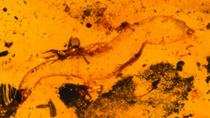
Oldest Known Male Gecko Found Preserved in Kachin Amber

Carinasquama rusmithii. Image credit: Daza et al., doi: 10.3897/sjp.145.188227.

Paleontologists have described a new genus and species of ancient gecko found in two pieces of amber from the 99-million-year-old (mid-Cretaceous) amber deposits in Kachin province, Myanmar.

The post Oldest Known Male Gecko Found Preserved in Kachin Amber appeared first on Sci.News: Breaking Science News.

  •  

385-Million-Year-Old Amber Found in China

The Middle Devonian Hujiersite amber. Image credit: Lu et al., doi: 10.1126/sciadv.aeh1266.

Paleontologists in China have discovered what they say is the earliest confirmed pieces of amber ever found, a fossilized resin roughly 385 million years old (Middle Devonian epoch) that predates the previous record-holder by about 65 million years.

The post 385-Million-Year-Old Amber Found in China appeared first on Sci.News: Breaking Science News.

  •  

The First Ancient Protein Analysis of Homo Naledi Has Revealed a New Burial Mystery

A team of researchers examined 23 teeth from at least 20 individuals from South Africa‘s Rising Star Cave system and found no evidence of the male-specific Amelogenin-Y protein.

A recent study, published in Cell, marks the first ancient protein analysis of Homo naledi and raises the possibility that the Dinaledi Chamber may contain predominantly, or possibly exclusively, female individuals. If confirmed, Homo naledi would become the first known non-human species to practice sex-specific burial.

A Decade-Old Puzzle

Since its discovery in 2013, Homo naledi has puzzled paleoanthropologists. This extinct human relative lived between 335,000 and 241,000 years ago and combined ape-like anatomy with distinct human features. So far, Homo naledi remains have only been found in the Rising Star Cave system in South Africa.

For over a decade, scientists noted that the adult fossils from the Dinaledi Chamber were nearly identical, lacking the skeletal variation usually seen between males and females. The reason for this similarity remained unclear.

Proteins Where DNA Fails

To investigate further, a team from the University of York, the University of Copenhagen, and additional institutions analyzed the remains at the molecular level. They used a minimally destructive acid-etching method to extract protein fragments, or peptides, from the enamel of the 23 teeth.

“Unlike those found in other remains like bone fragments, proteins in tooth enamel are preserved because dental enamel—the hardest tissue in the human body—shields proteins from environmental contamination even for millions of years. This makes them ideal carriers of genetic information from deep time. Our study helps explain the long-standing mystery of why Homo naledi lacked significant variation; it’s probably because they could have all belonged to one sex,” said Palesa Madupe, who conducted the research during her postdoctoral fellowship at the University of Copenhagen.

A Missing Male Marker

The researchers specifically searched for Amelogenin-Y, a protein produced only by the male Y chromosome, but detected none of it in the sampled teeth. To make sure the results were not due to modern contamination, a specialized chemistry team at the University of York independently confirmed that the amino acids were genuinely ancient. The findings remained consistent.

“The lack of male markers within the group is truly fascinating. It is incredibly exciting to gain a window not only into the biology of our ancestors, but also into how they lived. These findings offer rare insights into a culture that has, until now, been difficult to access directly. Advances in ancient protein analysis are opening the door to a far richer and more nuanced understanding of ancient hominins,” said Dr. Marc Dickinson from the university’s Department of Chemistry.

Ritual or Genetic Fluke?

The findings allow for two possible explanations. One is cultural: if the chamber were reserved for females, it could indicate that Homo naledi practiced sex-specific, symbolic mortuary behavior, a trait previously thought to be unique to modern humans.

The second explanation is biological. The researchers note that a small, isolated Homo naledi population could have lost or mutated the Amelogenin-Y gene, leaving males without the protein marker typically used to identify male remains.

Either explanation could reshape what scientists know about Homo naledi. If researchers confirm the site as an all-female burial chamber, it would suggest that symbolic behavior emerged earlier in the human lineage than previously believed. On the other hand, a genetic change across the population would show how isolation can shape a species’ biology in ways that fossils alone cannot reveal. This study shows how ancient protein analysis from teeth can open new windows into human evolution.

Austin Burgess is a writer and researcher with a background in sales, marketing, and data analytics. He holds an MBA, a Bachelor of Science in Business Administration, and a data analytics certification. His work focuses on breaking scientific developments, with an emphasis on emerging biology, cognitive neuroscience, and archaeological discoveries.

  •  
❌