❌

Reading view

Explainer: What is bioprinting?

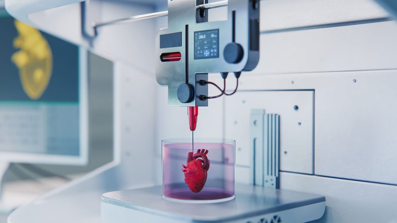
With the help of 3-D printers, even hobbyists can make model rockets or gaming accessories out of seemingly nothing. What if that printer could make new body parts, such as an ear, lung or skin? This may sound like science fiction. But scientists are already using living cells to “ink” prototypes of these tissues. 

This 3-D bioprinting uses the same basic strategy now used to print toys, sculptures, metal pieces or even basketballs. Specialized printers just swap out plastic or metal for living cells.

Actually, there’s a bit more to it than that. In most cases, you can’t use the same devices to make tissues. And you can’t just spew cells onto a surface, layer by layer, and expect them to link up into working organs. The cells have to be mixed with some soft, gel-like materials to make them unite with their neighbors.

Although still experimental, some devices have already printed skin, cartilage and even early-stage blood vessels. In 2025, researchers in Australia used an experimental robot to bioprint ink made from a patient’s own skin cells directly onto her burn wounds.

One of the doctors who pioneered the bioprinting of skin onto burn wounds describes how it’s done and why her team is excited about it.

It starts with the ink

In most 3-D printers, a resin, plastic filament or metal serves as the ink. Bioprinters instead use cells — and something to keep them alive.

“Bioinks are specialized soft gels that can have living cells mixed inside them,” explains Ryan Martin. He works on this tech at Virginia Commonwealth University, or VCU, in Richmond. Think of these inks like Jell-O: wobbly, soft materials that can hold their shape while protecting what’s inside.

Scientists make these hydrogels from materials such as alginate (AL-jin-ate) and collagen. Alginate comes from seaweed. Collagen is a protein found in skin and bones.

But bioinks contain more than just cells and hydrogel. That extra stuff — part of a complex matrix — helps the mix mimic the material that surrounds cells in the body, explains Ankita Pramanick. She’s a biomedical engineer at Utrecht University in the Netherlands.

Bioprinted tissue in space
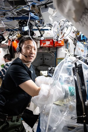
a photo of Jonny Kim working with bioprinted liver tissues on the International Space Station
NASA Johnson Space Center/Wikimedia Commons

NASA astronaut Jonny Kim treats bioprinted liver tissue on September 17, 2025, in a portable glove-bag on the International Space Station. These samples later went into another research device so researchers could study the effects of microgravity on blood vessel formation in engineered tissues.

What makes bioinks different from most other 3-D inks is that they host living cells. And those cells must survive the printing process, where they’ll get squeezed through nozzles and exposed to temperature changes. The matrix is designed with these challenges in mind.

After printing, the inked structures go into incubators, which are containers that maintain ideal conditions to help cells grow. There, the newly printed cells can mature, strengthen and grow together to form tissues — perhaps cartilage for joints or layered skin with blood vessels.

Printing living cells

Engineers have been developing several different ways to turn bioinks into tissue. Which one they choose depends on what they’re building.

Most times, a “bioink is pushed through a nozzle like toothpaste from a tube,” Martin explains. This process is known as extrusion. A machine controls how fast the ink comes out, building up a tissue layer by layer.

For example, to create skin tissue, you’d first print fibroblasts (FY-broh-blasts) — the cells that make up the inner layer of your skin. Later, you’d layer on keratinocytes (Kare-uh-TIN-oh-sites). These are the cells that make up our skin’s protective outer layer. Once printed, this combined structure matures within an incubator.

Extrusion works well for thicker, structured tissues. These include skin and cartilage. But it’s relatively slow. Squeezing cells through a nozzle can also stress them.

Inkjet bioprinting is faster and gentler. It sprays tiny droplets of bioink onto a surface in much the same way an office printer sprays ink onto paper. It works best with a very liquid matrix. That’s why it’s often used for thin cell layers, such as those that line the insides of blood vessels.

Another type of bioprinting creates entire structures at once instead of building them layer by layer. To start, a laser shines its light into a spinning tube of light-sensitive gel and cells. This solidifies them into the precise shape needed.

With this, scientists might print a small network of blood vessels all at once — complete with all its branches. It could take just seconds. And that’s important, because speed matters. The faster tissues are printed, the less time their cells spend outside their ideal environment.
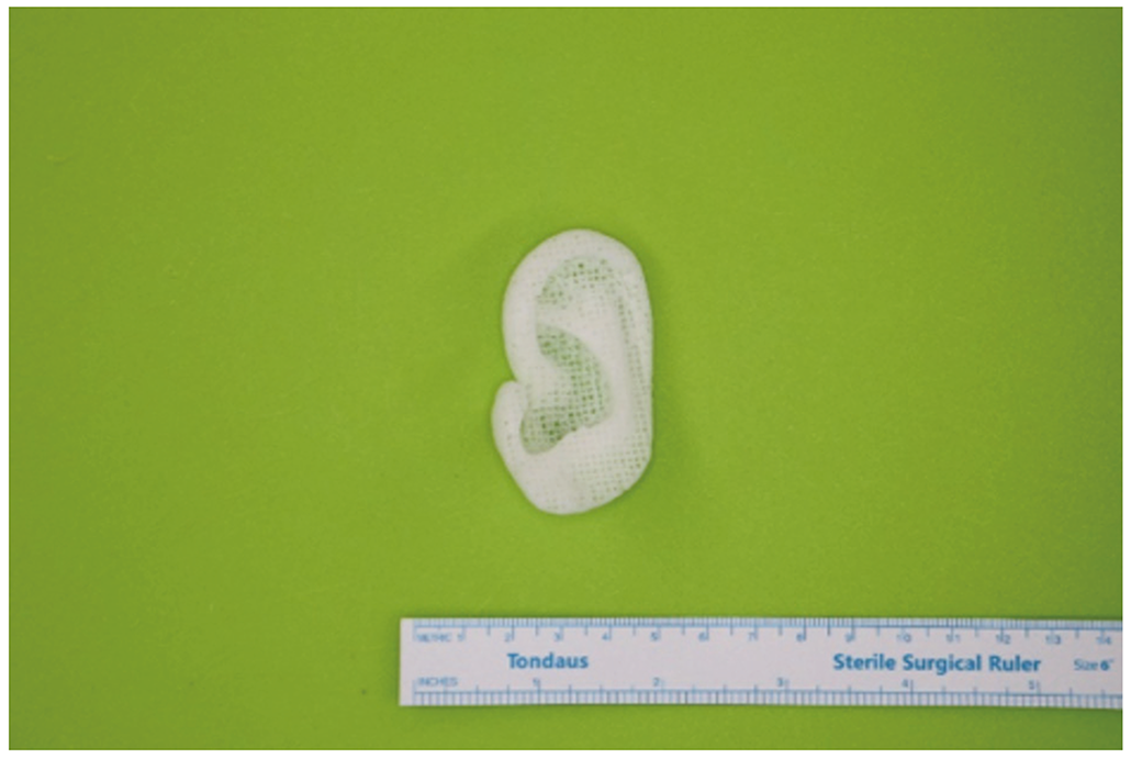
a photo of a 3-D printed ear implant made of a polymer
This 3-D printed ear implant was made from polycaprolactone (PCL), a biodegradable polymer. The implant is custom-designed to match a patient’s own ear and is shown next to a ruler for scale. The goal is to transition from plastic, as here, to implants made from living cells.M. Kim et al./Yonsei Medical Journal 2024 (CC BY-NC 4.0)

What’s being printed

In 2022, a patient received a 3-D–printed ear made from their own cells. In late 2025, surgeons successfully implanted a bioprinted cornea — the clear outer layer of the eye — into someone who had been legally blind.

Skin grafting is perhaps the most advanced application. A skin graft is a type of transplant where healthy skin is used to cover missing or injured skin. A 2025 trial in Australia tested bioprinted skin on burn patients. In a news conference with her doctor, the first patient to receive such a graft showed it had successfully worked on her leg.

Bone is trickier to print. Unlike skin or cartilage, it contains hard minerals mixed with living cells. Scientists have used 3-D printing to create bone scaffolds — frameworks that encourage the body’s own bone cells to grow and fill in the gaps. But no one has yet bioprinted actual bone with living cells in it for people.

Complex, fully-functioning organs such as hearts and kidneys are still a work in progress. Tissues thicker than a few millimeters (about a tenth of an inch) face a critical problem: Shortly after printing, their cells quickly start dying from a lack of oxygen and nutrients.

Heart of the matter
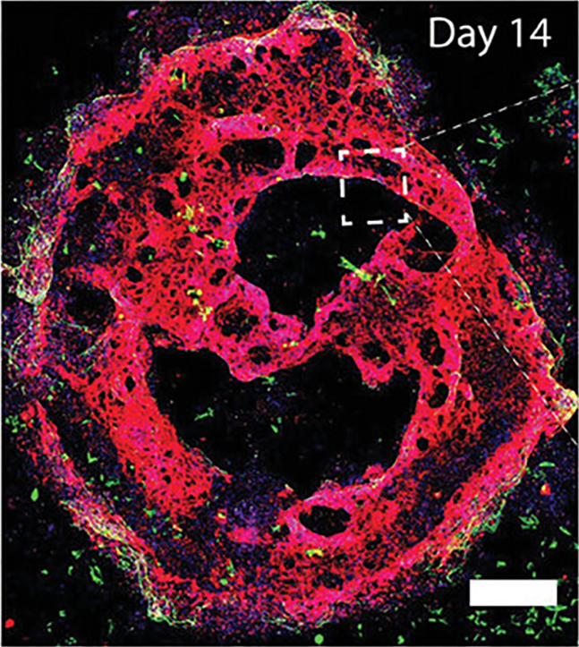
a section of bioprinted heartlike tissue under a microscope with false coloration
A Pramanick et al./Advanced Functional Materials 2025 (CC BY 4.0)

The image shows bioprinted heartlike tissue after 14 days in an incubator. The different colors help show the position of different types of cells inside the tissue.

The missing piece: blood vessels. These are needed for bringing oxygen to cells deep inside thick tissues and carrying away wastes. Without that oxygen and waste removal, cells will die. That’s why building blood vessels into tissues is the biggest hurdle standing in the way of fully printed organs.

The problem comes down to diffusion — the movement of molecules from areas of high concentrations to low concentrations. Oxygen can diffuse only a few millimeters into tissue. In thicker tissues, cells in the center would suffocate before oxygen could reach them. Imagine a big, densely populated city with no roads. Food trucks can’t bring in supplies. Garbage trucks can’t haul away waste.

Scientists have tried ways to avoid this problem. One method uses sacrificial inks. These serve as temporary placeholders. The inks are printed in the shape of vessels, then tissue is printed around them. Once the tissue solidifies, the sacrificial ink dissolves. In its place sit hollow channels through which blood can flow.

Scientists are also looking for a way to print blood vessels directly into tissue as it’s being built up.

Do you have a science question? We can help!

Submit your question here, and we might answer it an upcoming issue of Science News Explores

Moving forward

This short overview describes what bioprinting is, how advanced it’s become and where it might go.

In September 2025, researchers made a promising advance with a 3-D bioprinting system they’re calling GRACE. ”This technology enables the printing of blood vessels directly around [the printed tissue itself],” explains Pramanick at Utrecht. She works in the lab that’s developing GRACE.        

Here’s how it works. A computer-vision system “sees” where cells are located. It then asks artificial intelligence to figure out the best pattern of blood vessels to deliver oxygen-rich blood to all those cells. In seconds, bioprinting then creates pipe-shaped structures to begin delivering that blood.

Speed matters enormously here. Traditional methods might take hours to print vessel networks layer by layer. Because GRACE does it in seconds, cells spend little time without oxygen.

Full organs remain years away. Forming stable, treelike structures in different sizes — from big arteries down to the tiniest vessels known as capillaries — to seamlessly work with a patient’s blood-vessel system remains a big challenge, notes Martin at VCU. As bioinks and printing speeds improve, the prospect of integrating printed tissues with blood vessels into the body appears ever nearer. Today, the question is no longer if 3-D bioprinting can create replacement organs — it’s when.
  •  

Scientists made clocks from an atomic nucleus

For the first time, scientists have used an atomic nucleus as a clock.

Clocks need to keep a steady rhythm. (Think of the swinging pendulum in a grandfather clock.) The new clocks are based on how light interacts with an atomic nucleus — the collection of protons and neutrons at the center of an atom. This technique could allow scientists to make clocks that tick more precisely than any before.

The world’s most precise timepieces are already made using atoms. Existing atomic clocks rely on their electrons. Clocks based on atomic nuclei might perform even better. Now, two teams of scientists have finally made the first nuclear clocks.

This tech is still at an early stage. So nuclear clocks don’t yet tick more precisely than atomic clocks. But these new clocks can already test physics in novel ways.

A research team used one of these clocks to search for dark matter. That’s an unidentified substance that makes up much of the universe. The team describes its new clock — and the search — in a paper submitted June 3 to arXiv.org. This nuclear clock didn’t find any dark matter. But it seems to be more sensitive to some types than atomic clocks. In that search, “we’re already outperforming all of the atomic clocks,” reports Thorsten Schumm. He’s a physicist on the project at Vienna University of Technology in Austria.

“This is an outstanding result,” says Victor Flambaum, who did not take part in the work. A theoretical physicist, he works at the University of New South Wales in Sydney, Australia. The new feat should spur more progress, he says. “This is only the first step. [The] race for building super-accurate nuclear clocks just started.”

Do you have a science question? We can help!

Submit your question here, and we might answer it an upcoming issue of Science News Explores

The clocks’ laser ‘pendulum’

Nuclear clocks have the potential to weigh in on other weird physics, too. That’s why they’re “one of the most actively pursued frontiers,” says Shiqian Ding. He’s a physicist at Tsinghua University in Beijing, China. Ding’s team made a nuclear clock based on a technology similar to Schumm’s. They described this second clock in a paper submitted June 7 to arXiv.org. (Neither paper has been peer reviewed.)

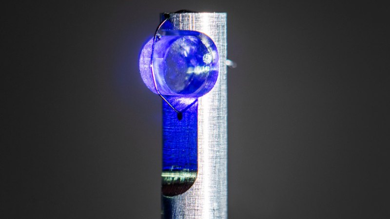
Both clocks contain crystals of calcium fluoride. Embedded in those crystals are atoms of thorium-229, a radioactive isotope. That thorium is key. In the entire periodic table, it’s the only element whose atomic nucleus can be used to make a clock.

Scientists hit that thorium-229 with a laser. The wiggling electromagnetic waves of laser light acted like a clock’s swinging pendulum. Thorium’s role was to make sure that the frequency of those waves didn’t change. This kept the “ticking” of these clocks from slowing or speeding up.

The way this works is based on quantum physics, the branch of science that describes atoms and similarly tiny stuff. According to quantum physics, a nucleus can hold only certain amounts of energy. That gives each nucleus a set of energy levels. A jump between two particular energy levels of a given atom always takes the same amount of energy.

So the laser’s frequency was locked to a jump between energy levels in the thorium nucleus. Only the right frequency of light will make that jump take place. Scientists used the jump to readjust the laser. They did this over and over to keep the ticks steady.

Thorium-229 is the only atomic nucleus that has an energy jump of a size that will be initiated by a laser. So it’s the only one that can be used to make a clock.

Although scientists have previously gotten close to making nuclear clocks, this readjustment step had never been done before.

“This was the final missing step before calling it an actual clock,” says Lars von der Wense, who also was not involved with the research. A physicist, he works at Johannes Gutenberg University Mainz in Germany. With improvements to lasers and crystals on the horizon, he says, nuclear-clock technology is expected to advance rapidly.

Improving on atomic clocks

Nuclear clocks have been hotly anticipated. Compared with atomic clocks, they’re less sensitive to stray electromagnetic fields that can throw them off. And they can be made out of solid materials. The atoms in atomic clocks must be suspended in a cumbersome vacuum chamber. These traits have scientists hoping for more portable, robust clocks.

And atomic nuclei respond to different forces than electrons. Electrons mainly are subject to electromagnetic forces. In contrast, the strong nuclear force holds protons and neutrons together. That opens up new possibilities for study.

Numbers called fundamental constants determine the relative strength of those forces. Comparisons of an atomic clock to a nuclear one could be used to check if those numbers really stay constant over time.

It’s been a long wait — almost a quarter century — since scientists first dreamt of a thorium nuclear clock. But “I have always been optimistic about the success of this project,” says Ekkehard Peik. He’s a physicist at the National Metrology Institute in Braunschweig, Germany. Peik is one of the scientists who proposed the idea for such a clock and was a coauthor with Schumm on a paper describing the new clocks.

After initially slow progress, researchers have made rapid advances in recent years. Now, Peik says, “a great deal of interesting research … is only just beginning.”

  •  

Songs prep unhatched finches’ brains for a hot world

Like many parents, zebra finches sing to their young. But some parental songs seem to prepare the baby birds for hot weather, even before they leave the egg.

Zebra finches live in sun-crisped Australian woodlands. On hot days, adults make a rapid, peeping “heat call.” It kicks off genetic changes in the brains of their unhatched babies. The tune acts like a weather forecast. It prepares them for the broiling conditions on the outside of the eggshell.

Researchers described their new findings June 11 in the Journal of Experimental Biology.

A decade ago, behavioral ecologist Mylene Mariette was part of a team that made a discovery. Exposure to heat calls in the egg, shortly before hatching, changed how chicks of these zebra finches (Taeniopygia guttata) dealt with high temperatures. They grew more slowly. They also preferred warmer places to nest. In all, they seemed better able to handle heat. 

But one mystery remained: How could hearing a simple song trigger all this?

Singin’ for the brain

Mariette, of Deakin University in Waurn Ponds, Australia, teamed up with neuroscientist Julia George. George works at Clemson University in South Carolina. The pair wanted to know if those songs might trigger changes in the brain’s hypothalamus. This region plays a big role in controlling the birds’ metabolism and responses to heat. 

Hear the finch’s heat-induced call

This high-pitched, rapid peeping is the “heat call” of Australian zebra finches. It spurs their unhatched babies’ brains to change how they move blood around. This might reduce the hatchlings’ risk of heat stroke — and lasts the birds’ entire life.

“Some of the consequences of [the call] are permanent. The birds will choose warmer nest sites as adults if they are exposed to heat calls as embryos,” says study author Julia George. “And they will produce more offspring in those warmer environments.”

AUDIO: M. MARIETTE

The researchers raised the eggs of zebra finches at a consistent temperature. Then they separated them into two groups. They exposed about half of these eggs to playbacks of an adult’s heat call. The other half heard a different call for a few days. Afterward, the team removed the embryos from the eggs and euthanized them.

Researchers cut out a small sample of the hypothalamus and extracted some of its RNA. This is a messenger molecule made based on DNA. It carries genetic information to the protein-making parts of the cell. The researchers planned to compare the levels of different RNAs. This should show how certain genes ramp up or down.

The team expected the heat calls would trigger changes in hormone-regulating genes, George recalls. Instead, the songs reduced the activity of genes that control how the brain’s blood vessels squeeze. The researchers now think this helps the chicks brains’ shed heat.

“It’s important that the temperature of the brain is kept cool, even in hot temperatures,” George says. Modifying blood flow there could protect against dangers such as heat stroke.

The calls didn’t change how the hypothalamus works, in general. Instead, only a select set of systems in it seemed to change. These systems are vulnerable to high temperatures. Only about 2 percent of the RNA in the hypothalamus showed this response to the heat calls.

The heat calls could be “like a little weather forecast,” George says, that allows finches to fine-tune their brain’s development. And this might leave them “better suited to the environment right after they hatch,” she adds.

Ready for a warmer world

Other types of yet-to-hatch birds have also shown sound-triggered changes. Yellow-legged gulls warn each other of predators. Those exposed in the egg to these warning calls grew more slowly. After hatching, they also were more likely to crouch when they heard those calls.

Alexandra Cones wonders if there are other genetic and cellular ways for animals to adapt to heat. Cones is an evolutionary behavioral ecologist at Ludwig Maximilian University. It’s in Munich, Germany. She did not take part in the new finch study. But her own research has shown that adult house sparrows can change how their chicks’ metabolisms respond to temperature.

Cones’s team never uncovered exactly how this happens. But, she says, the zebra finch data “forces us to consider a broader range” of possibilities for how such a flexibility evolved.

Zebra finches may not be alone in their brain-bending musical abilities, George concludes. Perhaps there’s a whole playlist of heat-busting birdsongs yet to be discovered.

Do you have a science question? We can help!

Submit your question here, and we might answer it an upcoming issue of Science News Explores

  •  

Scientists Say: Alloy

Alloy (noun, “AL-oy”)

An alloy is a metal-based mixture. Familiar examples of alloys include steel, bronze and brass. Some alloys are metal-metal mixtures. Others mix metals with nonmetals.

Alloys offer strong, flexible support for skyscrapers, jewelry and more. By adjusting the recipe of an alloy, metalworkers can fine-tune its properties. That way, they create the perfect material for the job.

Take steel. This alloy contains the elements iron and carbon. Iron is metallic element. Adding carbon to it strengthens the metal. The result is a versatile metal alloy that is strong, flexible and resilient. That makes it perfect for making everything from a thin knife blade to scaffolding for our tallest buildings.

An alloy made only of carbon and iron is called carbon-steel.  But often, chemists add other elements to fine-tune the properties. For instance, a little bit of chromium added to the mix creates stainless steel. This upgraded version greatly increases steel’s resistance to rust. It works because the chromium combines with oxygen in the air. Technically, this is how rust forms. But in the case of stainless steel, the chromium “rust” forms a barrier that does not flake away like iron rust. This barrier protects the iron — which, on its own, is a very rust-prone metal.

Pure silver is a metal element. But sterling silver is an alloy. For millennia, silver’s beautiful, rust-resistant gleam has inspired craftsmen to create jewelry. In the modern age, silver’s high electrical conductivity makes it an important metal for electronics. However, silver is rarely used on its own. Like gold, silver is a “soft” metal. Jewelry made from pure silver would warp out of shape. So, metalworkers blend in a bit of copper or some other metal for added strength. Almost all silver jewelry is made from alloys. To be considered “sterling” silver, the alloy must contain 92.5 percent silver.

In a sentence

Alloys made from rare-earth metals can create super magnets, superconductors and more.

Check out the full list of Scientists Say.

  •  

Fog — it’s alive! 

A bank of fog might seem gloomy. But that mist may carry an unexpected community of environmental helpers. Ground-dwelling clouds can house bacteria that clean the air, a new study finds.

Microbes can find air a harsh place, notes Kathleen Weathers. Temperatures can fluctuate broadly. And even in fog, there can be a high risk of drying out. A fog scientist, Weathers works at the Cary Institute of Ecosystem Studies in Millbrook, N.Y. But, she adds, fog hosts many things needed for those microbes to “make a living.” These include moisture, nutrients and comfortable temperatures.

Yet until now, scientists hadn’t known microbes could actually live there, says Weathers, who did not take part in the new work.

It took Thi Thuong Thuong Cao, a chemist who loves hiking on foggy mountainsides, to show microbes not only can live in fog, but also grow and reproduce.

Collecting clouds 

As a chemist, Cao wondered what might be inside fog droplets. At first, she focused on their chemistry. Soon, though, she teamed up with other scientists to see what microbes might hitchhike in fog — and what they might be doing.

At the time, Cao was a graduate student at Arizona State University in Tempe. Part of her team traveled to Pennsylvania, where another team member was based. The area had great spots to collect fog.

This team focused its hunt on a special type of low-lying cloud: radiation fog. It forms at night when the ground cools and then lowers the air’s temperature. Cooler air can’t hold much water. So it sheds the excess into the air as small droplets, creating fog.

Radiation fog doesn’t roll in. Instead, it forms in place. So any microbes should be local, not ones carried in from elsewhere.

Over about two years, the team collected samples of water from 32 “fog events.” They used a device called a cloud-water collector. It pulls air into a chamber containing strings.

scientific equipment set on grass on during fog
Researchers assembled this setup in a Pennsylvania field to collect fog samples. They were looking to capture not only fog but also anything that might be living in the airborne water droplets.T.T.T. Cao

As fog condenses on the strings, it forms droplets that drip into a bottle below. The scientists collected up to 200 milliliters (about 7 ounces) of fog water per event. That’s roughly the volume of a juice box. They also collected samples of the air in between the fog droplets. They trapped similar air samples before and after fog events.

The fog samples housed a multitude of microbes. In fact, fog water is about as densely populated with microbes as ocean or lake water. When examined under a microscope, many fog-water microbes seemed to have been dividing when they were collected. That led the researchers to conclude the microbes had been growing in the fog.

Next, the team extracted the fog-dwellers’ RNA, a type of genetic information. Cao’s team compared these types of genetic maps to those in a database on known microbes. That helped them identify the fog’s inhabitants.

An average of 188 unique genetic patterns showed up in each fog event. Each represented a unique type of bacteria. The communities of microbes present were similar across all fog samples. But they differed quite a bit from those found in air without fog.

Cao and her team shared their findings in mBio on May 22.

Do you have a science question? We can help!

Submit your question here, and we might answer it an upcoming issue of Science News Explores

Turning pollution into food 

Their analyses showed Methylobacterium was the most abundant microbe in fog water. It accounted for up to half of all microbes present.

To grow in fog, the team reasoned, those Methylobacteria must have found something in it to eat. This microbe is known to consume carbon-containing chemicals, such as formaldehyde.

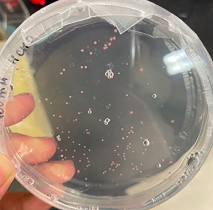
a petri plate showing some of the colonies of bacteria sampled from the fog
Researchers grew colonies of Methylobacterium in this dish, feeding them only formaldehyde, a toxic chemical. “[T]hey have [a] pink pigment,” says Thi Thuong Thuong Cao. “They’re really cute.”T.T.T. Cao

Formaldehyde is a common and toxic air pollutant. Breathing it can cause harm. Some formaldehyde comes from natural sources, such as rotting plant materials. Human activities, such as construction and burning fossil fuels, also release it. It’s even one of the chemicals that makes cigarette smoke harmful.

Methylobacteria are well suited for dealing with this chemical, Cao says. “They can survive formaldehyde at a concentration higher than other bacteria [can].” They break it down hundreds of times faster than other microbes, too.

Her group measured formaldehyde in freshly collected fog water. Over several hours, levels of the chemical fell surprisingly fast.

The researchers also isolated Methylobacterium from fog water and then grew it on its own. These bacteria were able to quickly remove formaldehyde when exposed to an artificial fog water containing the chemical.

It disappeared so quickly, in fact, that the bacteria couldn’t have used it all for growth. The microbe may break the chemical down as well as eat it, the team concludes. Such a strategy might keep formaldehyde from reaching toxic levels.

This all seems to confirm Methylobacterium can remove formaldehyde from fog. The researchers don’t yet know how the bacteria might affect air quality. But their new data now point to fog as a potential ecosystem.

Fog can transport water and microbes across a landscape. These data now suggest that as it does, it may play a role in shaping the chemistry of the environment.

  •  
❌