The technology behind flying cars is continuing to advance, with companies developing vehicles that combine elements of traditional cars, helicopters, electric aircraft, and — in Tesla's case — rockets. Many of these designs use electric vertical-takeoff-and-landing technology, which allows them to lift off and land without a runway by using multiple rotors or propellers to generate lift. Advances in battery technology, autonomous flight systems and computer-assisted navigation are helping to make these vehicles seem more feasible. But challenges remain, including questions surrounding passenger safety, affordability, and the need for regulationsand laws and infrastructure for the widespread use of these vehicles.
Tesla says production of its new "flying car" is scheduled for 2028, but the company is notorious for not meeting its proposed deadlines. So do you think flying cars could actually be available to the public within the next five years? Or are we still a long way from turning this science fiction fantasy into reality? Cast your vote in our poll, and share your thoughts in our comments section.
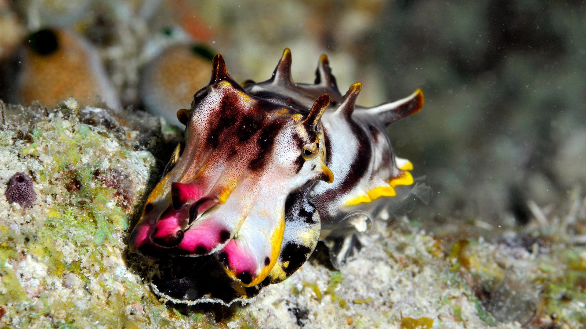
Like their cephalopod cousins the octopuses, cuttlefish are masters of disguise.
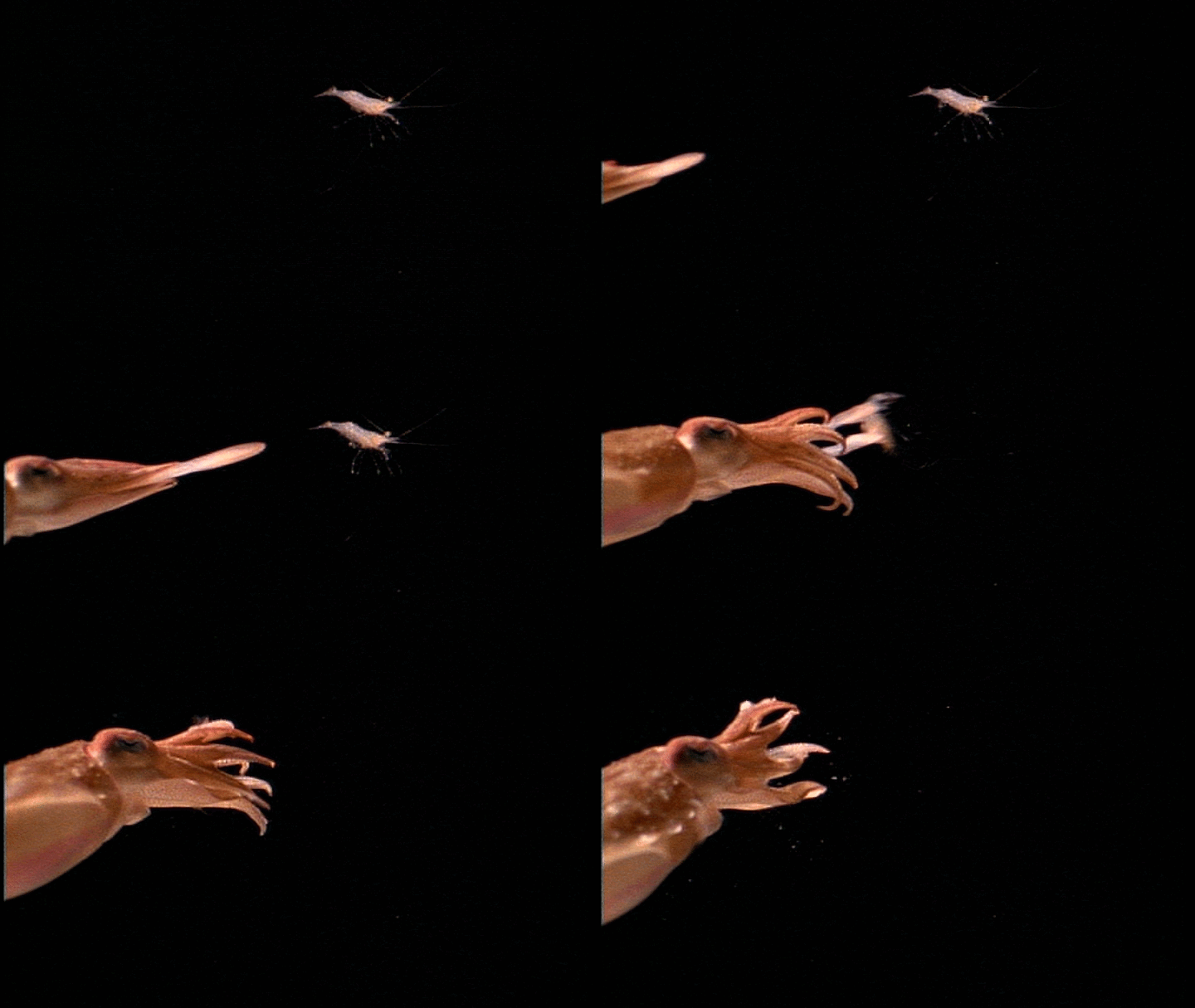
As they hunt, these marine predators send waves of color across their skin. The rainbow display stops prey in their tracks, granting the cuttlefish just enough time to shoot out two specialized hunting tentacles at lightning speed and snatch them into range of their deadly beaks.
But can cuttlefish actually mesmerize prey with their colorful skin changes? Or is something fishier at play?
Experts told Live Science that cuttlefish are ingenious hunters, but that there's likely an even more mysterious process than hunter to prey hypnosis going down on the seafloor.
A calculated hunter
Cuttlefish are visual hunters, using their large, complex eyes to locate and track potential meals. In general, their strategy involves a slow, stealthy approach, said Bret Grasse, the manager of cephalopod operations at the Marine Biological Laboratory in Woods Hole Massachusetts.
"The cuttlefish will slowly approach the prey so as to not alert the unsuspecting shrimp, fish or crab," Grasse told Live Science in an email.
During their approach, cuttlefish often put on a showy display, producing moving bands across their body. These displays are produced by a combination of light-reflecting pigment cells known as iridophores, leucophores and chromatophores. These cell types contract and expand to produce specific colors and patterns, all within milliseconds.
Some may wonder whether this trick can mesmerize prey, but scientists don't think that's the case.
Hypnosis, defined as a mental state of increased relaxation and altered awareness, is thought to be a uniquely human psychological state. There is very little research on whether fish can be hypnotized, but a 2005 study found that pushing a steady stream of water through the snapper (Chrysophrys auratus) put them in an immobile state similar to being hypnotized. Another study looked at sedative hypnotic drugs like benzodiazepines and barbiturates and their effects on zebrafish, finding that the fish had molecular receptors for these drugs, which helped render them immobile. However, whether fish can be visually put into a state like hypnosis has yet to be directly studied.
And while some animals, such as chickens, can be induced into trance-like conditions of tonic immobility, an involuntary, temporary state of unresponsiveness that happens when humans or animals experience extreme trauma, it's unlikely that the cuttlefish's color changes "hypnotize" their prey, Connor Gibbons, the cephalopod facility manager at Columbia University told Live Science. Instead, some researchers believe that the color show serves as an ingenious disguise.
As waves of different colors ripple down the cuttlefish's body, the shape of the animal becomes less obvious to its prey, so they may not see the cuttlefish as a threat to flee from, Gibbons said.
"It's only part of something looming at them, which they're like, 'I don't know if you are a predator? Are you just passing waves? What are you?'" Gibbons said. "And then by that time, it's too late."
A cuttlefish uses its tentacles to capture a shrimp (Image credit: Oxford Scientific Films via Getty Images)
Once they get close enough, the cuttlefish shoots their two long, specialized feeding tentacles from small hidden pouches near the animal's mouth at their prey.
"The closest thing I can compare it to is the tongue of a chameleon extending out to grab an unsuspecting insect," Grasse said. "Cuttlefish feeding tentacles will fire out in the blink of an eye before the prey can react and escape."
Then, the prey is pulled toward the cuttlefish's mouth, pierced by its sharp, chitinous beak and injected with venom. The use of the venom helps the cuttlefish reduce their energy usage when hunting "as the cuttlefish does not want to use unnecessary energy and struggle against the prey while it's trying to eat," Grasse said.
However, the notion that cuttlefish flash colors to hide their body shape is only one idea of why cuttlefish perform these strobing color displays. Other researchers, including Grasse, describe them as a way to distract their prey.
"Essentially, they are trying their best not to alarm the prey to their presence," he said. By breaking up the size of their bodies, cuttlefish can convince their prey they're not as large or alarming as they may seem.
Whether the light shows are for disguise or distraction still needs parsing out, Gibbons said, and scientists also need a better understanding of how the cuttlefish controls its deadly dermal display. "The jury is still out on exactly what is happening," he said.
How much do you know about octopuses? Find out with our octopus quiz!
Recent research challenges a long-standing idea about how the brain develops: Rather than arising from a single source, the brain may emerge from two distinct populations of cells, the study finds.
"The brain appears as one organ," said study co-author Kyle Loh, an associate professor of developmental biology at Stanford University. "However, we discovered that the front part of the brain and the back part of the brain come from different sources, which are pushed together to create the appearance of a single organ," Loh told Live Science in an email.
The findings, published in September in the journal Nature Neuroscience, not only challenge established models of how the brain forms but may also explain why researchers have struggled to grow certain types of brain cells in the laboratory. The scientists say the work could offer new ways to investigate diseases that affect the brainstem, the "stalk" that connects the brain to the spinal cord.
Two developmental journeys
The human brain is divided into three main regions — called the forebrain, midbrain and hindbrain — that perform different but interconnected functions.
The forebrain, which includes the cerebral hemispheres, is involved in higher-level cognitive processes, such as language, planning and abstract thinking. The midbrain, the uppermost part of the brainstem, connects the forebrain and the hindbrain and acts as a key relay center for sensory information and movement control. The hindbrain — which includes the bottom of the brainstem, along with other structures — also helps to coordinate movement, as well as regulate essential functions such as breathing, sleep and heart rate.
Historically, the hindbrain has been difficult to study. One reason is that scientists have faced persistent challenges in generating functional hindbrain neurons in laboratory conditions.
To help understand why, Loh and his team looked at the early stages of embryonic development in lab mice. They focused on gastrulation, the point when cells begin to organize into structures that will later form different organs. The researchers identified two distinct populations of immature cells that would go on to give rise to nervous system tissue, including the brain.
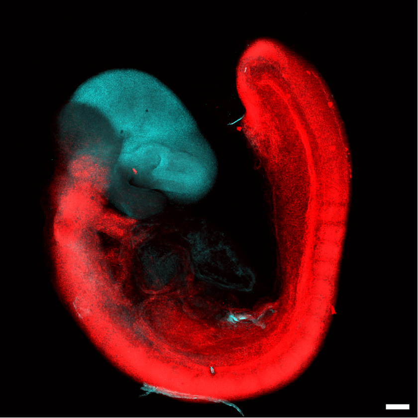
One cell population expressed a gene called Otx2, which helps to direct the early development of the forebrain and midbrain. The second group of cells expressed the gene Gbx2, which is involved in the development of the midbrain and hindbrain.
This photograph shows a 9.5-day-old mouse embryo and depicts the two separate developmental origins of the brain. The forebrain and part of the midbrain (blue) express the Otx2 gene, while the hindbrain and the rest of the midbrain (red) express Gbx2. The red cell population also extends into the spinal cord. (Image credit: Stanford Medicine)
The functions of these genes were already known, but their distinct expression in these two groups of cells was revealed by the new research. The cell populations remained distinct as they developed into full-fledged nervous tissue, rather than overlapping.
The team also examined chromatin, the molecular structure that packages DNA tightly inside the nucleus and helps regulate which genes a cell can use. The two cell populations show different chromatin configurations, the team found, which further supports the idea that they follow separate developmental pathways.
That may help to explain why scientists' attempts to grow hindbrain neurons have been largely unsuccessful.
"Previous attempts to make hindbrain neurons likely tried to coax forebrain and midbrain progenitors into hindbrain cells, which our study shows is not possible," study co-author Rayyan Jokhai, a graduate student in Loh's lab, said in a statement.
Why it matters
Using the information uncovered in their experiments, the researchers successfully guided human stem cells to transform into functional hindbrain motor neurons, which are involved in controlling movement. Specifically, these cells produced proteins characteristic of the hard-to-grow hindbrain regions that help control muscles of the face and those involved in swallowing.
In several neurological diseases, including spinal muscular atrophy and amyotrophic lateral sclerosis, motor neurons deteriorate and affected patients can lose their ability to swallow, speak and breathe independently.
"We cannot sample living motor neurons from a patient's hindbrain to watch how these diseases attack these neurons," Loh said. "Growing these hindbrain motor neurons in a dish gives us a way to investigate these diseases." Looking forward, the team aims to use their findings to study these diseases and pinpoint new treatments.
The new findings could also reveal something fundamental about the evolution of vertebrate animals.
The study authors investigated whether the two developmental pathways were unique to humans or reflected a much older evolutionary arrangement. They turned to previously published data on other species — including macaques (Macaca), zebrafish (Danio rerio) and chickens (Gallus gallus domesticus) — and again found two distinct cell populations expressing the two key genes.
And even in acorn worms — a marine invertebrate whose evolution diverged from vertebrates about 550 million years ago — they again found these two distinct groups of cells.
"The Stanford discovery establishes that the deep ancestry of the vertebrate brain is fundamentally modular," said Anna Di Cosmo, a professor of biology at the University of Naples Federico II in Italy who wasn't involved with the study. "The developmental separation between the anterior lineage (forebrain/midbrain) and the posterior lineage (hindbrain) is traceably ancient," Cosmo told Live Science in an email.
Loh's team speculates that these developmental trajectories seen in the womb may suggest that the two cell populations once belonged to two separate nervous systems. "Later in evolution, these two nervous systems may have been pushed together to give rise to the appearance of one integrated brain," Loh said.
However, the study doesn't reveal exactly when the two pathways arose or establish how they integrated. "It is important to emphasize that this is only an idea, and it might change as scientists learn more," Loh said.
See how much you know about the most complex organ in the human body with our brain quiz!
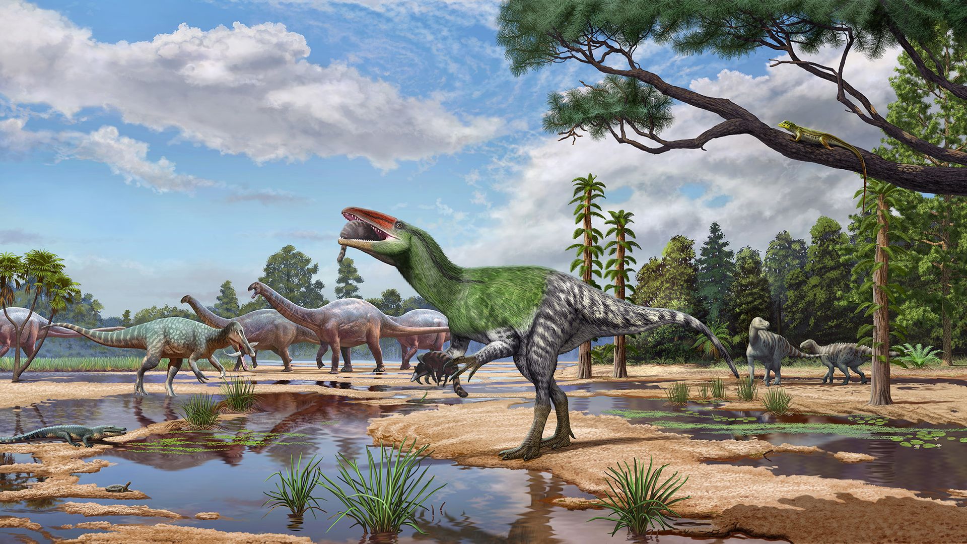
A fossil that sat in a museum collection for over a century is a previously unknown species of predatory dinosaur, researchers have found. The discovery could help scientists solve a longstanding mystery about how some of the Cretaceous period's most formidable hunters spread around the globe.
The dinosaur lived on what is now the Isle of Wight in southern England about 120 million years ago. It belonged to a group of two-legged carnivores called megaraptors, which are related to tyrannosaurs. But unlike tyrannousars, megaraptors are thought to have relied on their powerful arms and large curved claws to attack prey and defend themselves.
The newly identified species was described in an Oct. 14 study in the journal Royal Society Open Science. It is the first megaraptor identified in Europe, filling an important gap in the fossil record of a group whose evolutionary history remains poorly understood.
"It is a real mystery that this fossil was overlooked for so long, sitting in a drawer for nearly 100 years," study first author Cassius Morrison, a researcher in the Department of Earth Sciences at University College London (UCL) said in a statement. "Lots of bones are found on the Isle of Wight — who knows what else is waiting to be discovered."
Unconquered female warrior dinosaur
The discovery of the megaraptor began in 1924, when an amateur fossil collector discovered a dinosaur skull fragment on the Isle of Wight. The specimen eventually entered the collections of London's Natural History Museum, but it was largely forgotten.
Recently, researchers revisited the fossil as part of a study on dinosaur braincases. To identify the animal, the researchers used micro-CT scanning to examine the fossil's internal structure and build detailed 3D digital models of the space that once held the dinosaur's brain.
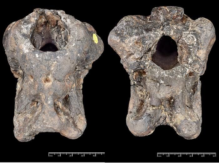
The researchers then compared the animal's anatomy with that of other meat-eating dinosaurs, including tyrannosaurs, spinosaurs and other theropods (the group of two-legged, mostly meat-eating dinosaurs). Several features differentiated the unknown specimen, including an almost oval opening where the spinal cord entered the skull and an unusual, hourglass-shaped ridge at the back of the braincase.
The skull of the newly identified theropod dinosaur, Invictibellatrix amberina. The hole is where the brain would have sat. (Image credit: The Trustees of the Natural History Museum, London)
Computer-based evolutionary analyses placed the fossils within the Megaraptora family tree, supporting the idea that the skull represented a never-before-seen species. The researchers named the dinosaur Invictibellatrix amberina, combining the Latin words "unconquered" and "female warrior" for the genus name, while amberina refers to amber found in the rocks associated with the fossil.
Megaraptors lived between roughly 125 million and 66 million years ago and varied considerably in size. While early members were relatively small, some later species became enormous predators. Maip macrothorax, discovered in Argentina, may have reached up to 33 feet (10 meters) long and weighed as much as an elephant. Based on comparisons with related dinosaurs, I. amberina was much smaller, likely around 10 to 13 feet (3 to 4 m) long and standing roughly 5 feet (1.5 m) tall. The researchers estimated it weighed around 440 to 660 pounds (200 to 300 kilograms), making it one of the smallest known megaraptors.
"It was not the apex predator of its time, but for humans this dinosaur would have been more dangerous than a T. rex," Morrison said. "We would have been perfectly sized prey, about a third of their body weight. They definitely would have chased us."
Although the newly described fossil doesn't preserve the animal's arms or claws, its closest relative suggests that its claws may have measured around 4 to 8 inches (10 to 20 centimeters) long. The researchers also suggested I. amberina may have been feathered, though that feature can't be determined directly from the specimen.
A missing link in the dinosaurs' globetrotting
Scientists have long suspected that megaraptors originated in Asia before spreading to other parts of the world, eventually becoming the dominant predators across the southern supercontinent Gondwana, which included present-day Africa, South America and Antarctica. But a lack of European fossils left an important gap in verifying this migration route.
The new study's evolutionary models suggest that I. amberina descended from Asian megaraptors that dispersed into Europe around 130 million years ago. From there, members of the group may have moved through what is now Africa and further into Gondwana via temporary landbridges and temporary land connections across the shallow Tethys Ocean.
"This new species is the first proof we have that megaraptorans were also present in Europe and that they reached Gondwana this way rather than island-hopping further south," study co-author Charlie Scherer, a doctoral student at UCL Earth Sciences, said in the statement.
However, the specific route these animals took over millions of years is still unknown. The researchers also cannot yet explain why megaraptors disappeared from northern areas, but changes in climate or competition with other predatory dinosaurs may have contributed. More research on other megaraptor fossils like I. amberina could help uncover more clues.
"Work like this shows the exceptional value of working with historical museum collections where there are still many discoveries to be made," study co-author Paul Barrett, a researcher of dinosaur evolution at the Natural History Museum in London, said in the statement.

 Login