
❌

Normal view

Siloton secures £8.4m to speed development of portable retinal imaging system

16 October 2026 at 14:30

UK medical technology start-up Siloton has been awarded an £8.4 million contract to accelerate the development of its portable eye imaging system. Based on optical coherence tomography (OCT), the company’s GiraffeOCT device generates detailed retinal images for detection and monitoring of a range of treatable eye diseases.

OCT is a non-invasive optical technique that creates high-resolution, depth-resolved images of biological tissue. While OCT is the clinical gold standard for diagnosing retinal diseases, existing systems are large and expensive and typically only available in hospitals and optometry clinics. By using photonic chip technology to shrink the bulky optical components onto a single coin-sized chip, Siloton is developing an easy-to-use, handheld binocular-style system.

The contract, from Innovate UK, will massively accelerate development of both the in-clinic GiraffeOCT system and the at-home version, which will have built-in automation and guidance so that users can reliably image themselves. “The contract will reduce the time to market for the medical device by one to two years,” says Alasdair Price, CEO of Siloton. “It will also allow us to grow the company, bringing in new skills around manufacturing and regulation, both of which are critical to the next stage of the business.”

Continuing clinical trials

In 2024, Siloton was the first commercial organization to demonstrate sub-surface imaging of a human retina, using its first-generation Akepa OCT photonic chip. The following year, the company trialled its device with patients for the first time, imaging their retinas to identify the presence and progression of age-related macular degeneration (AMD). Testing continued as the system developed, with a second cohort of patients imaged earlier this year.

In addition, the Project Ginan consortium in Australia has purchased a GiraffeOCT research system and independently evaluated its retinal imaging performance with a group of healthy volunteers.

Over the course of the two-year Innovate UK contract, Siloton will work with Sunderland Specialist Eye Hospital in the UK to perform clinical trials of the in-clinic device, giving patients access to the technology during its development. “These trials will be based around the prototype for the full medical device, providing additional post-processing and improved ergonomics compared to our research-use only product line,” Price explains.

The funding will also support the development and internal testing of Siloton’s second-generation CheetahOCT system, a research-use device that will measure different layers of the retina, expanding the number of conditions that it can detect. CheetahOCT incorporates the company’s second‑generation BlueJay chip – the same architecture used in the system that Siloton is building for astronauts – and will be available from early 2027.

“The image performance will be significantly improved, increasing the number of biomarkers that it can be used to measure. This means you will be able see more of the eye and in finer detail,” Price tells Physics World. “CheetahOCT will also be much more compact, as we continue to miniaturize other parts of the system that fall outside the photonic chip.”

The post Siloton secures £8.4m to speed development of portable retinal imaging system appeared first on Physics World.

Una carta de 1571 descubierta en Budapest confirma la autenticidad del misterioso Códice Rohonc y revela que el escriba que lo redactó estaba aterrorizado

14 October 2026 at 19:06
Hay manuscritos medievales que nadie sabe leer y hay uno, el manuscrito Voynich, que se ha vuelto casi una marca registrada del misterio. Guardado en la Universidad de Yale y fechado en el siglo XV, ha servido de inspiración para novelas y teorías de todo tipo. Pero existe otro libro que los especialistas suelen colocar […]

Miniature microprobe enables internal imaging of smaller blood vessels than ever before

23 September 2026 at 09:00

Cardiovascular disease is the leading cause of death globally, with atherosclerotic disease – caused by the build-up of fatty plaques in the arteries – responsible for around three-quarters of these deaths. Modern intravascular imaging allows us to see these fatty plaque build-ups in detailed cross sections, revolutionizing atherosclerosis diagnosis and treatment planning. A research team in China is pushing this imaging technique to smaller blood vessels and higher resolution than ever before. Their approach could even enable imaging of the cerebrovasculature – the network of tiny vessels within the brain.

Intravascular optical coherence tomography (IV-OCT) uses near-infrared light emitted and received at the end of a catheter to image the walls and texture of the blood vessel. It works by comparing the wavelength and phase offset of reflected light from tissues with reference light reflected from a mirror, with 360° sensor rotation generated by electromagnetic motors.

The two most common designs of OCT catheters are proximal (where rotational force is generated outside of the body) and distal (with rotational force generated by a small motor inside the catheter). However, proximal systems suffer from friction-induced rotational distortion in smaller vessels, while distal scans are blocked from full 360° vison by wire artefacts.

Size is a major limitation in IV-OCT imaging. The catheters traditionally used are around 2 mm in diameter and perform optimally in vessels of 10 mm in diameter – medium/large arteries in the body. This diameter is limited by the size of electromagnetic motors needed to induce the sensor rotation and the wiring of these motors.

In this latest work, the researchers – led by Dawei Wu at Nanjing University of Aeronautics and Astronautics and Rui Liu at Nanjing University Medical School – show that it is possible to make a tiny 0.55 mm diameter probe, optimal for imaging 2 mm vessels, whilst retaining 360° field-of-view. The piezoelectric microprobe achieves this small size, and resolves both the rotational distortion and limited field-of-view difficulties, by exploiting a functionally different miniature drive mechanism.

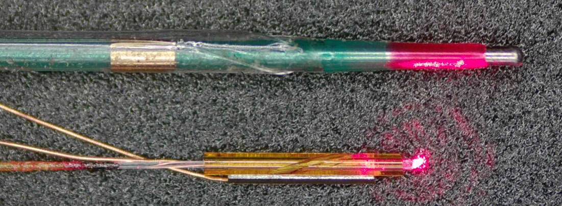
A piezoelectric micro probe and a commercial balloon dilatation catheter
Dimension comparison The piezoelectric micro-optical probe (bottom) and a commercial balloon dilatation catheter (top). (Courtesy: Dawei Wu, Nanjing University of Aeronautics and Astronautics)

The microprobe uses a single-phase AC circuit and piezoelectric crystal to vibrate a glass tube. A 10° groove in the glass translates this longitudinal vibration into torsional vibration, thus generating elliptical motion of the lens in a similar way to a crank and slider. When AC voltage is applied, the piezoelectric crystal expands and contracts and the lens rotates, thereby producing the required optical scanning.

Device testing

First author Boquan Wang and colleagues first tested whether their probe reduces rotational distortion by imaging metal tubes arranged around a small, curved vessel. The probe was able to scan the tubes at 50 revolutions per second while maintaining angular deviation of just 1° – a significant improvement compared with the 9° found in traditional proximal IV-OCT catheters. They note that this test also demonstrated the probe’s full 360° field-of-view and that the single-phase voltage required to rotate the lens meant that the wiring was minimal.

The researchers confirmed the probe’s ability to navigate and image small vessels via successful tests on leaf microveins, a vascular stent and ex vivo pig vessels. In an important milestone in assessing the suitability of the probe for imaging the cerebrovascular system, the probe successfully traversed through a full-scale human vascular model to reach the middle cerebral artery.

Finally, to verify the efficacy of the probe in identifying pathological lesions, the team compared images of human plaques acquired by the probe with histological analyses. The OCT and histology findings agreed well in identifying locations of plaque rupture sites and regions high in collagen fibres.

Why does this matter?

By putting a novel miniature rotational motor directly inside the probe and powering it with a single-phase AC circuit, the team created a probe that’s smaller than traditional IV-OCT probes and eliminates rotational torsion artefacts while achieving full 360° imaging.

Importantly, the improved catheter navigation could allow access to high-resolution images of the structure of arterial walls in smaller, more curved vessels than is currently possible clinically. This opens the door to assessment of plaque pathology and planning of stent positions in the heart–brain system.

“Although the probe would need more development and testing to be used clinically, intravascular OCT could one day give physicians a much closer look at what is happening inside the small arteries of the brain,” says Wu in a press statement.

The researchers describe the microprobe in Biomedical Optics Express.

The post Miniature microprobe enables internal imaging of smaller blood vessels than ever before appeared first on Physics World.

❌