
❌

Normal view

Open-source software tracks wrist bone motion in 3D and 4D CT scans

9 September 2026 at 18:20

In a development that could reshape how scientists study the mechanics of human movement, a team of biomedical engineers and computer scientists has unveiled an open-source software tool capable of tracking the tiny, intricate bones of the wrist as they glide and rotate during motion. The new tool, called Hierarchical 3D Registration, or 3DH, is a module within the SlicerAutoscoperM extension of the widely used 3DSlicer computing environment. Described in a study published in BioMedical Engineering OnLine, the software was validated against previously established methods using both sequential three-dimensional computed tomography, or 3DCT, scans of living participants performing thumb tasks and dynamic four-dimensional CT, or 4DCT, images of cadaveric wrists moving through flexion and extension. The results demonstrate that the open-source approach can match the accuracy of independent, previously published methodologies while offering researchers everywhere a free and reproducible path to quantifying skeletal kinematics.

The wrist has long been one of the most challenging joints for biomechanists to study. It contains eight small carpal bones, including the scaphoid and lunate, along with the radius of the forearm and the metacarpal bones of the hand, all articulating in a tightly packed arrangement that allows the remarkable range of motion humans rely on every day. Understanding how these bones move relative to one another, a field of study known as arthrokinematics, is essential for unraveling the causes of wrist instability, osteoarthritis, ligament injuries and the long-term outcomes of surgical repairs. Yet measuring those movements in living, moving joints has historically required either invasive techniques, labor-intensive manual analysis or proprietary software that few laboratories can access or verify.

Quantitative tracking of skeletal structures in vivo, with both efficiency and high temporal resolution, matters to a wide range of questions in biomechanical research, and the authors of the new study argue that the field has been held back by a shortage of accessible, semi-automated registration tools. Image registration, the computational process of aligning one three-dimensional image volume to another, lies at the heart of motion tracking from CT data. When a bone moves between two scans, its position and orientation change, and a registration algorithm must find the transformation that best overlays the bone’s image from one time point onto the next. Done well, this yields precise measurements of rotation and translation. Done poorly or inconsistently, it introduces errors that can swamp the small movements of carpal bones, which may shift by only a few millimeters and a few degrees during normal function.

The new 3DH module addresses this problem with a hierarchical strategy for registering bones across sequences of image volumes. The researchers presented and evaluated the software using two distinct classes of data drawn from previous studies. The first consisted of sequential 3DCT datasets of human participants performing various thumb tasks, in which the radius, the first metacarpal and the trapezium were the target bones. The second consisted of dynamic 4DCT datasets capturing simulated flexion-extension motion in cadaveric wrists, providing time-resolved, three-dimensional image volumes of the scaphoid and lunate in motion. By testing the tool on both static sequential scans and true dynamic imaging, the team was able to demonstrate that 3DH is not limited to a single imaging paradigm but can serve as a unified tracking solution across modalities.

To determine whether the new open-source tool could be trusted, the investigators compared the arthrokinematics computed with 3DH against values that had been previously calculated using independent approaches for the same 3DCT and 4DCT datasets. The statistical framework for this comparison was the Bland-Altman analysis, a standard method for assessing agreement between two measurement techniques. Rather than simply correlating the two sets of results, Bland-Altman analysis quantifies the mean bias, the systematic difference between methods, and the 95 percent limits of agreement, the range within which most individual differences between the methods are expected to fall. This approach is particularly well suited to method-comparison studies because it reveals whether a new technique introduces systematic error and how widely individual measurements might deviate from those of the established gold standard.

The performance figures reported in the study are striking. For the 3DCT data, in which the radius, first metacarpal and trapezium were tracked, the mean bias for the helical angle, a single-axis description of three-dimensional rotation, was just minus 0.06 degrees, with 95 percent limits of agreement ranging between minus 1.87 degrees and 1.74 degrees. Translation measurements fared nearly as well: the mean bias was 0.12 millimeters, with limits of agreement spanning from minus 2.23 millimeters to 2.00 millimeters. In practical terms, the open-source tool reproduced the rotational and translational measurements of the earlier methods to within roughly two degrees and two millimeters at the extremes, differences small enough to preserve the biological signal in most wrist kinematics experiments.

The dynamic 4DCT results told a similar story for the carpal bones. Tracking the scaphoid and lunate through simulated flexion-extension, 3DH produced mean biases of the Euler angle rotations, which decompose three-dimensional rotation into three orthogonal components, of less than 0.77 degrees about any direction. The widest 95 percent limits of agreement ranged between minus 7.11 degrees and 8.65 degrees. For translations, mean biases were less than 0.5 millimeters in any direction, with the widest limits of agreement ranging between minus 2.36 millimeters and 1.43 millimeters. The wider limits of agreement for rotations in the dynamic data likely reflect the added challenges of time-resolved imaging, including motion blur and the smaller, more complexly shaped carpal bones, but the near-zero mean biases indicate that the tool does not systematically drift away from established values.

Crucially, all target bones were successfully tracked in both the 3DCT and 4DCT datasets, meaning the hierarchical registration algorithm converged on correct solutions in every case tested. This robustness matters because registration algorithms can occasionally fail, locking onto incorrect alignments or diverging entirely, and such failures typically demand time-consuming manual intervention. A semi-automated tool that reliably succeeds across diverse bones, motion tasks and imaging protocols removes a significant bottleneck. Manual bone tracking in CT volume sequences can take hours per bone per trial, and the labor multiplies rapidly in studies involving multiple participants, multiple bones and dozens of motion cycles. By automating this process within a validated framework, 3DH promises to accelerate the pace at which wrist biomechanics data can be collected and analyzed.

The choice to build the module inside 3DSlicer, and specifically within the SlicerAutoscoperM extension, is a deliberate strategic decision with implications well beyond the wrist. 3DSlicer is a free, open-source platform for medical image computing and visualization with a large international user base spanning research, clinical and educational communities. By embedding 3DH in this ecosystem, the developers have made the tool immediately available to any laboratory already working with 3DSlicer, complete with the platform’s established infrastructure for loading DICOM data, segmenting anatomical structures and visualizing results. The authors note that the ability to track bones from both 3DCT and 4DCT data sources, with agreement to prior methodologies, demonstrates that the 3DH approach is a capable tool that could promote collaboration and consistency of results through open-source software.

Consistency is a quietly urgent issue in biomechanics. When different laboratories use different proprietary tools with undisclosed algorithms, it becomes difficult to distinguish true biological differences between study populations from mere methodological discrepancies. Open-source registration changes that calculus: the code can be inspected, verified, extended and reused, and results from different institutions become directly comparable because they are produced by identical algorithms. This transparency also lowers the barrier to entry for research groups in settings that cannot afford commercial licenses, potentially broadening the global community capable of conducting rigorous skeletal kinematics research.

The study itself emerged from a collaboration spanning several institutions. The team included Joseph J. Crisco, Amy M. Morton and John D. Holtgrewe of the Department of Orthopedics at The Warren Alpert Medical School of Brown University and Rhode Island Hospital; Cesar Lopez, Andrew Thoreson and Kristin D. Zhao of the Assistive and Restorative Technology Laboratory and the Department of Physical Medicine and Rehabilitation at the Mayo Clinic in Rochester, Minnesota; and Jean-Christophe Fillion-Robin and Beatriz Paniagua of Kitware Inc. in Clifton Park, New York. The involvement of Kitware, a company known for its leadership in open-source scientific computing, underscores the blend of academic rigor and software engineering expertise required to produce a research tool robust enough for widespread distribution. Funding was provided by the National Institute of Arthritis and Musculoskeletal and Skin Diseases of the National Institutes of Health under award numbers AR059185, AR078924 and AR071338.

The implications of this work extend naturally to the future of musculoskeletal imaging. As newer CT technologies, including photon-counting detector systems, deliver ever-finer spatial resolution and as dynamic 4DCT becomes more widely available, the volume of image data available for kinematic analysis will grow dramatically. Tools like 3DH, which are fast, semi-automated and validated across modalities, will be essential for converting that imaging bounty into quantitative biomechanical knowledge. For patients with wrist disorders, the downstream promise is equally tangible: better measurements of how carpal bones actually move in living joints can inform more accurate diagnoses, improved surgical planning, more realistic computational models of joint loading and ultimately better-designed treatments for conditions ranging from scapholunate instability to thumb-base arthritis. With the tool now openly available in the 3DSlicer environment, researchers around the world can begin putting it to work.

Subject of Research: Open-source kinematic tracking of the small bones of the wrist in sequential 3DCT and dynamic 4DCT images using Hierarchical 3D Registration

Article Title: Kinematic tracking of the small bones of the wrist in sequential 3DCT and dynamic 4DCT volume images using open-source Hierarchical 3D Registration, a module within SlicerAutoscoperM

Article References: Crisco, J. J., Morton, A. M., Lopez, C., Thoreson, A., Zhao, K. D., Holtgrewe, J. D., Fillion-Robin, J.-C., & Paniagua, B. (2026). Kinematic tracking of the small bones of the wrist in sequential 3DCT and dynamic 4DCT volume images using open-source Hierarchical 3D Registration, a module within SlicerAutoscoperM. BioMedical Engineering OnLine. https://doi.org/10.1186/s12938-026-01601-x

Image Credits: AI Generated

DOI: 10.1186/s12938-026-01601-x

Keywords: Computed tomography, 3DCT, 4DCT, Bone tracking, 3DH, 3DSlicer, Image registration, Wrist kinematics, Arthrokinematics, Open-source software, SlicerAutoscoperM, Bland-Altman analysis

Physicists Find Useful Energy Hiding in Quantum “Waste Heat”

8 September 2026 at 18:23
Heat Engine in Driven Dissipative Quantum SystemA new theoretical approach helps connect quantum physics with classical thermodynamics by clarifying what counts as heat and useful work in tiny quantum machines. When a machine is reduced to a single atom and particles of light, even basic concepts such as heat and useful work become difficult to define. This problem sits at the [...]

Researchers shrink double-slit experiment to atomic scale

8 September 2026 at 09:00
Shrinking Young’s double-slit experiment
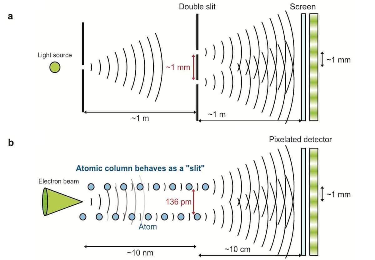
Shrinking Young’s experiment (a) The classical double-slit experiment using light (slit spacing ~1 mm, propagation distance ~1 m). (b) Schematic of atomic-scale interference in which a pair of neighbouring silicon atomic columns act as a “double slit” for a focused electron probe (spacing 136 pm; crystal thickness ~10 nm; distance to detector ~10 cm). (Courtesy: Koudai Tabata/University of Tokyo)

For the first time, physicists in Japan have carried out an atomic-scale version of the double-slit experiment. Using an ultraprecise electron beam channelled along two adjacent columns of atoms in a crystal of silicon, Naoya Shibata and colleagues at the University of Tokyo observed clear interference fringes in electrons scattered by the atoms: fringes that persisted even at high temperature, thanks to correlations in the atoms’ thermal vibrations.

The double-slit experiment is perhaps the most iconic demonstration in quantum mechanics: showing how a single particle’s wavefunction interferes with itself as it passes through two slits at once, with a tangible impact on where the particle is ultimately observed.

In practical research, this effect is now routinely harnessed in electron, neutron and atom interferometers. By observing the interference fringes that it creates, researchers can extract valuable information about both the quantum matter wave and whatever object is doing the diffracting.

Recently, physicists have attempted to push double-slit interferometry all the way down to the atomic scale, replacing conventional slits with crystal lattices. When a moving electron encounters an atom in a crystal, it scatters across a range of angles – producing an outgoing spherical wave that’s mathematically identical to the diffraction pattern from a classical slit.

Since atoms in a crystal sit at such well-defined separations, any neighbouring pair of them could, in principle, act as a double-slit interferometer. In reality, however, the many repeated unit cells in a crystal mean the resulting interference fringes are washed out, making it difficult to isolate the signal from a single atomic pair.

To address this challenge, Shibata’s team used the ultraprecise probe of a 4D scanning transmission electron microscope: an instrument designed to capture electron diffraction patterns with a pixellated detector. Fired into a pure silicon crystal at just the right orientation, the microscope’s precision let the researchers target two adjacent columns of aligned silicon atoms, separated by just 136 pm. As a result, the electron wave overlapped both columns, and no others.

“Our key idea was to use these two atomic columns as the two slits,” describes team member Takehito Seki. “By channelling the probe along both columns at once, we turned them into two coherent scattering sources – effectively making the crystal itself into an atomic-scale double-slit interferometer.”

At this scale, Shibata and Seki’s team created electron interference fringes some seven orders of magnitude smaller than in Thomas Young’s original double-slit experiment with light, first carried out in 1801.

Just as in a larger-scale experiment, these patterns encoded information about the crystal and the electron beam. But at this scale, the researchers could also detect the signature of individual phonon modes in the columns. Rather than destroying coherence as uncorrelated atomic motion would in a classical picture, these correlated vibrations between the two atomic columns kept them moving in sync, preserving the interference.

“In a which-path picture, relative motion of the two atomic columns makes the two electron pathways more distinguishable and reduces the interference, whereas correlated in-phase motion preserves coherence,” Shibata explains. As a result, distinct interference fringes remained visible across temperatures ranging from 300 to 900 K.

The team’s results already point to how an atomic-scale double-slit experiment could offer new ways to measure phonon correlations directly between single pairs of atoms. But as Shibata explains, the possible applications could stretch much further.

“It also opens a route to probing local lattice dynamics at the scale of individual atomic bonds, including at interfaces and defects, where local lattice dynamics can strongly influence thermal transport,” he says. “In the long run, such atomic-scale insight could help inform better thermal management strategies for future chip technologies.”

The study is published in Nature.

The post Researchers shrink double-slit experiment to atomic scale appeared first on Physics World.

Meet the 2026 Ig Nobel Prize winners

3 September 2026 at 18:00

It's that time of year again, when we learn which lucky scientists are among the winners of the Ig Nobel Prizes. This year, the prizes honor research on designing the perfect splash-free urinal; using mosquito proboscises to "necroprint" tiny nozzles; studying composition rates of buried cotton underwear; and the aerodynamics of a healthy nose-blow, among other highlights.

Established in 1991, the Ig Nobels are a good-natured parody of the Nobel Prizes; they honor “achievements that first make people laugh and then make them think.” The unapologetically campy awards ceremony features miniature operas, scientific demos, and "24/7 lectures," whereby experts must explain their work twice: once in 24 seconds and the second in just seven words.

Acceptance speeches are limited to 60 seconds. And as the motto implies, the research being honored might seem ridiculous at first glance, but that doesn’t mean it’s devoid of scientific merit. In the weeks following the ceremony, the winners will also give free public talks, which will be posted on the Improbable Research website.

Read full article

Comments

© YouTube/Improbable Research

First-of-a-Kind Experiments Demonstrate Effects of Einstein’s Gravity in the Quantum World

3 September 2026 at 13:29

An international research team has announced the first-ever successful observation of gravity’s effect on a falling quantum object, demonstrating a connection between gravity and the quantum world first predicted by Albert Einstein.

The research team said the result does not unify quantum mechanics and gravity, nor does it prove that gravity is a quantum phenomenon.

However, the researchers said their study offers experimental support that Einstein’s equivalence principle, a fundamental concept at the heart of the famed scientist’s theory of gravity, remains consistent with matter in the quantum world.

Unifying Einstein’s Theory of Gravity with Quantum Mechanics

When physicists want to describe nature, they rely on two distinct sets of rules. Einstein’s theory of gravity explains the mechanics of large-scale phenomena like stars and galaxies. Conversely, quantum mechanics helps scientists characterize the behavior of extremely small objects. Still, finding a scientific bridge uniting the two sets of rules has proven elusive.

“These two pillars of modern physics have so far eluded all attempts at a unified theoretical framework,” explained Professor Ron Folman from Ben-Gurion University of the Negev and the lead author of the new study.

Einstein did, however, identify one mechanism where the two worlds meet. Called the equivalence principle, the concept says that gravity should effectively disappear for an observer in free fall. For example, NASA simulates zero-gravity environments by flying astronauts in a plane, making successive rising and falling parabolic arcs. During the short periods of falling, the astronauts briefly experience ‘weightlessness” due to the equivalence principle.

quantum gravity
Above: The 2D MOT apparatus which feeds the science chamber with cold atoms. At the centre is a glass cell held under vacuum between coils producing magnetic fields. The atoms go into the science chamber through the tiny hole that may be seen in the centre of the yellow (reflecting) surface (Image Credit: Or Dobkowski).

Curious if the same phenomena would apply to the quantum world, the research team, which included Nobel Prize-winning physicist Professor Sir Roger Penrose, designed experiments. According to a statement announcing the research, the team hoped their setup would allow them to measure a “distinctive change in the quantum properties of atoms as they fell under gravity.”

Experiments Show Gravitational Pull Affecting Quantum Objects

To search for gravity’s effect in the quantum world, the researchers created clouds of rubidium atoms cooled to just above absolute zero. The researchers used a specially designed atom chip that allowed the team to manipulate the ultracold rubidium atoms.

Next, PhD student Or Dobkowski and colleagues used microwave pulses to place the atoms in quantum superposition. This unique quantum state lets each atom travel down two paths at once.

Using tiny electrical wires embedded on the specialized chip, the researchers generated tunable magnetic fields. The researchers said the field’s precision tunability allowed them to apply an upward force to one half of the atomic wave. For this experiment, the team created a magnetic field at the precise strength needed to counter gravity, effectively holding that half of the wave stationary relative to Earth.

After suspending the first half of the wave, the team used a magnetic pulse to lift the second part. Then, the team switched to a state almost unaffected by the magnetic field, allowing it to fall under gravity.

Once the second half completed its fall, the team used magnetic fields to reunite the two halves. When reunited, the two halves of the atomic wave interfered with each other. The researchers said that interaction allowed them to “measure the tiny difference in quantum phase accumulated while one was falling and the other was held still.”

“The phase measured in the new experiment is the same as the one predicted when Einstein’s principle is applied to such a quantum wave,” they explained.

“One of the Most Fundamental Questions in Physics”

When discussing the results, the research team said their result should not be construed as proof that gravity is quantum. The results also do not unite gravity and quantum mechanics. However, Folman noted, “this complex experiment gives more hints as to how such a unification may be achieved.”

Study co-author Professor Vlatko Vedral from the University of Oxford’s Department of Physics agreed, while also noting that their experimental results successfully demonstrated Einstein’s equivalence principle’s consistency with quantum mechanics, which alone provides an experimental connection between quantum physics and Einstein’s theory of gravity.

“This experiment pushes quantum mechanics into one of its most intriguing frontiers, gravity, and shows that, once again, its predictions hold,” Professor Vedral explained.

The professor also noted the experiment’s combination of hard experimental data with “a far-reaching theoretical interpretation, about one of the most fundamental questions in physics: How can gravity (described by Einstein’s theory of relativity) and quantum theory be unified into one understanding of the universe?”

The study “Observation of the quantum phase of free fall and the consistency with the equivalence principle” was published in Science Advances. 

Christopher Plain has spent the last six years as Associate News Editor and Head Science Reporter at The Debrief. Follow and connect with him on X, learn about his novels at plainfiction.com, or email him at christopher@thedebrief.org.

❌