
❌

Normal view

Comprehensive Microbiome Analysis Uncovers Reliable Colorectal Cancer Biomarkers

24 June 2026 at 16:26

For decades, the scientific community has recognized the gut microbiome—the intricate ecosystem of bacteria and microorganisms residing within the human intestine—as a potential key player in colorectal cancer development. Yet, the complexity and variability of microbiome data have often obscured clear, reproducible links. Now, an ambitious new study spearheaded by the Mi-EOCRC consortium, which includes pioneering teams at EMBL Heidelberg, has transcended these obstacles by analyzing an unprecedented breadth of data to unearth a universal microbiome signature associated with colorectal cancer.

Garnering insights from an extensive meta-analysis of 27 studies with over 6,700 gut microbiome profiles, the researchers uncovered microbial patterns that remain consistent across diverse populations, sequencing methodologies, and age groups—illuminating a microbial footprint intrinsic to colorectal cancer itself. Their integrative approach not only encompassed stool samples but also expanded to 906 intestinal tissue specimens, revealing striking similarities between microbes colonizing tumors and those present in fecal matter.

This landmark research fundamentally shifted microbiome science by developing innovative computational tools capable of harmonizing heterogeneous datasets produced by different sequencing technologies. Such methodological rigor was critical in overcoming previous barriers of reproducibility that have long plagued microbiome studies. Central to their approach was a bespoke machine learning algorithm adept at categorizing microbiomes along a continuum from ‘healthy’ to ‘cancer-like,’ yielding a quantifiable score that can be leveraged across disparate human gut microbiome datasets, including those derived from dietary interventions.

This algorithmic precision allowed the consortium to isolate a colorectal cancer-associated microbiome signature that transcends traditional cohort boundaries. Intriguingly, the signature appears robust not only in late-stage disease but also in early-onset cases, dispelling prior uncertainty surrounding the microbiome’s role throughout the disease timeline. The discovery underscores the microbiome’s potential as an informative biomarker capable of reflecting the intricate disease ecology.

Moreover, by comparing tumor tissue microbial profiles with those detected in stool, the study illuminated how colorectal cancer’s microbial hallmarks are mirrored across sample types. Although the microbial signature was more readily detectable in advanced tumors or those proximal to the rectum, early-stage lesions exhibited a detectable microbial presence as well. This finding deepens our understanding of tumor-microbiome crosstalk and raises critical questions regarding how microbial communities might influence tumor progression via metabolic and signaling pathways.

Despite these advances, the researchers caution that pre-cancerous adenomas—often the target of preventive strategies—remain notoriously elusive in stool-derived microbiome analyses. The microbial alterations associated with adenomas were subtler and less consistent than those observed in full-fledged cancers. Sophisticated machine learning models specifically aimed at adenoma detection showed variable success across cohorts, highlighting the need for methodological refinement before such tools can make meaningful clinical contributions.

Diet emerged as another pivotal factor shaping cancer-associated microbial patterns. A compelling link was established between low dietary fiber intake and a heightened presence of the colorectal cancer microbiome signature. Conversely, dietary interventions increasing fiber consumption correlated with marked reductions in this cancer-associated microbial pattern. These findings reinforce the concept that diet—and particularly fiber—modulates the gut environment in ways that could impact cancer risk and progression, opening avenues for microbiome-focused preventive strategies rooted in nutrition.

Delving further into microbial taxonomy, the study provided high-resolution insights into Fusobacterium species, bacteria recurrently implicated in colorectal cancer. The researchers delineated notable differences among subspecies, with Fusobacterium nucleatum subsp. animalis exhibiting consistent cancer enrichment worldwide, whereas others showed distinct geographic distributions, predominantly in Asian populations. This nuanced understanding challenges the oversimplification of grouping all Fusobacteria uniformly and underscores the importance of subspecies-level resolution for future microbiome research and therapeutic targeting.

Open science was a cornerstone of this monumental undertaking. By consolidating thousands of publicly available microbiome profiles, the researchers achieved a scale of analysis previously unattainable, setting a new standard for evidence synthesis in microbiome studies. This collective data sharing facilitates reproducible science and paves the way for scalable, global approaches to understanding the microbiome’s role in human disease.

While the study stops short of proposing an immediate diagnostic application, it lays critical groundwork for the development of microbiome-informed clinical tools. Compared to existing non-invasive colorectal cancer screening tests like the fecal immunochemical test, microbiome-based classifiers are not yet on par for sensitivity or specificity. However, the reproducible microbial signature defined here could serve as a foundational reference for future machine-learning models, enabling improved risk stratification and early detection when integrated with other clinical markers.

The implications of this research ripple beyond colorectal cancer alone. The computational tools and conceptual frameworks unveiled may be applied to myriad disease contexts where the microbiome plays a role. Furthermore, the iterative feedback loop between dietary modulation and microbial signatures encourages a personalized medicine approach that incorporates lifestyle factors into disease management.

Finally, this research highlights the intricate dance between host, microbes, and environment that shapes disease landscapes. As microbiome science marches forward, studies like this exemplify the power of interdisciplinary collaboration, computational innovation, and open data stewardship to unravel complex biomedical puzzles, bringing us closer to novel interventions and preventive strategies that could transform patient outcomes worldwide.

Subject of Research: People
Article Title: Meta-analysis reveals microbiome signatures for colorectal cancer that are universal across onset age and sequencing method
News Publication Date: 24-Jun-2026
Web References: http://dx.doi.org/10.1016/j.chom.2026.05.030
Image Credits: Daniela Velasco/EMBL
Keywords: Colorectal cancer, Human microbiota, Gut microbiota

Hierarchical Endpoints, Win Stats in Geromedicine Trials

24 June 2026 at 15:17

In the rapidly evolving field of geroscience, the quest to translate scientific breakthroughs into tangible clinical benefits has encountered a formidable obstacle: the challenge of selecting trial outcomes that comprehensively reflect the multifaceted effects of geroprotective interventions while satisfying rigorous clinical and regulatory benchmarks. Despite the surge in research advancing our understanding of aging and potential therapies to extend healthspan, clinical trials often struggle to identify endpoints that are simultaneously robust, feasible, and accepted by regulators. This conundrum has impeded the pace at which promising discoveries transition from the bench to bedside.

Traditional endpoints in clinical trials for aging-related therapies have heavily relied on mortality as the gold standard outcome. Mortality is undeniably objective and resonates with regulatory agencies due to its clear clinical significance. However, using mortality as a primary endpoint is frequently impractical in geromedicine trials. The extended timelines required, the large participant populations needed, and ethical considerations make mortality a difficult measure for many intervention studies focused on aging. Given the complexity and prolonged nature of aging processes, mortality-based outcomes often limit the feasibility of clinical research into interventions designed to slow or reverse age-associated decline.

As an alternative, surrogate measures aimed at capturing the quality of extended life, often termed healthspan, have gained attention. Healthspan encompasses diverse dimensions such as physical function, cognitive performance, and biomarker profiles that collectively reflect an individual’s biological and functional status. These measures present a more practical and nuanced depiction of aging’s trajectory and the impact of geroprotective treatments. Nevertheless, healthspan-related endpoints are frequently regarded as “soft” or indirect, necessitating further validation to convince stakeholders of their reliability and clinical relevance. Their subjective nature and variability introduce challenges in affirming their predictive value and regulatory acceptance.

In response to these challenges, researchers have proposed an innovative methodological framework that seeks to harmonize the strengths of both mortality and healthspan measures while mitigating their respective weaknesses. This novel strategy employs hierarchical composite endpoints analyzed through a time-to-worst-event approach, leveraging win statistics to facilitate a pragmatic and scientifically grounded compromise. The essence of this methodology lies in comparing participant pairs within clinical trials based on a predefined, clinically justified hierarchy of events, prioritizing the most severe and objective endpoints such as mortality and major morbidity.

This hierarchical design assigns differing weights to outcomes positioned along a clinical severity spectrum. At the highest tier reside unequivocal events—death and serious clinical incidents—that carry unquestionable clinical importance and regulatory weight. Subsequent tiers integrate healthspan surrogates and biomarker data, contributing valuable subsidiary information that captures subtler yet meaningful intervention effects. Employing this layered approach enables researchers to reflect a fuller spectrum of aging-related outcomes without compromising the prominence of critical clinical endpoints.

Time-to-worst-event analysis serves as a robust statistical tool within this hierarchical framework. By focusing on the timing of the most severe event experienced by an individual, the methodology ensures that the earliest and most significant detriment determines the outcome classification, encompassing the patient’s clinical journey comprehensively. This time-sensitive lens captures not only whether but also when adverse clinical states manifest, providing a dynamic and nuanced perspective critical for aging-related trials, where the temporal dimension is essential.

Central to the proposed framework is the use of “win statistics,” a comparative metric allowing pairwise analysis of trial participants. This statistical method calculates wins, ties, and losses based on the clinical hierarchy, elucidating the overall treatment effect by aggregating these individual pairwise comparisons. The win statistic’s strength lies in its ability to synthesize multifaceted outcomes into a single interpretable measure while maintaining clinical meaning and relevance. Unlike conventional single-endpoint analyses, this approach acknowledges the multidimensional impact of geroprotective interventions.

Importantly, the selection and ordering of outcomes within this hierarchical system demand rigorous clinical and mechanistic justification. Consensus must be achieved among investigators, clinicians, and regulatory authorities to define which events take precedence and how ties are resolved. This transparent and collaborative process assures that composite endpoint construction reflects true clinical priorities and biological plausibility, bolstering regulatory confidence and facilitating trial approval and interpretation. Such an approach safeguards against arbitrary outcome weighting or biased interpretation.

The implications for the future of geromedicine clinical trials are substantial. By embracing hierarchical composite endpoints and win statistics, researchers can design more efficient trials that capture a broad spectrum of treatment effects within feasible timescales and participant cohorts. This methodology sidesteps the pitfalls of relying solely on mortality, while strengthening the evidentiary value of surrogate endpoints and biomarkers. Consequently, promising geroprotective interventions may undergo more straightforward, cost-effective evaluation, accelerating their clinical translation and potential public health impact.

This innovative design also opens pathways for more personalized assessment of intervention efficacy. By integrating multiple health domains and clinical events, trials can better reflect the heterogeneity of aging populations and diverse therapeutic mechanisms. The nuanced outcome assessment permits differential benefits and risks to be dissected, fostering precision geromedicine. Regulators and clinicians alike stand to gain from richer and more actionable data that truly capture quality-of-life improvements alongside traditional survival metrics.

Moreover, the potential regulatory acceptance of hierarchical composite endpoints could catalyze a paradigm shift in aging-related drug development. As the geroscience community demonstrates the validity and utility of such outcomes, regulators may become more amenable to approving longevity therapeutics supported by multifaceted endpoints rather than awaiting protracted mortality data. This regulatory evolution promises to invigorate investment and innovation in geromedicine, an area of growing importance in the context of expanding elderly populations worldwide.

While promising, this approach also entails challenges. It requires meticulous endpoint definition and standardization, robust statistical modeling expertise, and extensive stakeholder engagement to ensure that composite hierarchies are clinically meaningful and mechanistically grounded. Ensuring reproducibility and cross-trial comparability necessitates development of consensus guidelines and open data standards. Ethical considerations surrounding the relative weighting of life quality versus survival further complicate endpoint formulation and require ongoing discourse.

Nevertheless, the proposed hierarchical endpoint framework with win statistics embodies a timely and much-needed advancement addressing a fundamental bottleneck in geroscience clinical research. By bridging regulatory requirements with scientific insights into aging’s multidimensional nature, it lays a robust foundation for accelerated translation of geroprotective therapies. As the population demographic shift intensifies demand for effective aging interventions, such pioneering statistical and clinical innovation is pivotal in transforming geromedicine from promising science into practical medicine.

The work by Abdellatif, Kim, and Kroemer offers a compelling blueprint for the next generation of geromedicine trials. Their integrative approach thoughtfully balances objectivity, clinical relevance, and feasibility, paving the way for interventions that not only extend lifespan but also enhance the quality of those extended years. The advent of hierarchical composite endpoints guided by win statistics may ultimately revolutionize how aging therapies are evaluated, deployed, and embraced by healthcare systems worldwide, heralding a new era in age-related healthcare.

As this methodology gains traction, collaborative efforts across academia, industry, and regulatory bodies will be critical to refine, validate, and implement hierarchical endpoints in ongoing and future geroscience trials. Real-world application and longitudinal data will provide further insights to optimize outcome hierarchies and better reflect patient-centered benefits. Such collective progress holds the promise to unlock the full potential of geroprotective interventions, reshaping the landscape of aging medicine.

In essence, hierarchical composite endpoints and win statistic analysis emerge as a paradigm-shifting solution to the age-old dilemma of measuring meaningful outcomes in aging research. By embracing complexity and clinical priorities simultaneously, this approach captures the multidimensional health impacts of geromedicine interventions with rigor and pragmatism. The coming years will reveal how this innovation transforms clinical trial design and ultimately delivers on the long-awaited promise of prolonging both lifespan and healthspan.


Subject of Research: Geroscience clinical trial outcome measurement; hierarchical composite endpoints in geromedicine studies; win statistics for multidimensional aging-related outcomes

Article Title: Hierarchical endpoints and win statistics for geromedicine trials

Article References:
Abdellatif, M., Kim, Y. & Kroemer, G. Hierarchical endpoints and win statistics for geromedicine trials. Nat Aging (2026). https://doi.org/10.1038/s43587-026-01158-3

Image Credits: AI Generated

DOI: https://doi.org/10.1038/s43587-026-01158-3

UofL Scientists Reveal How Combining Fruits and Nuts with Specific Gut Microbes Promotes Intestinal Healing

24 June 2026 at 14:55

In a groundbreaking study poised to reshape the landscape of inflammatory bowel disease (IBD) treatment, researchers at the University of Louisville have elucidated the molecular mechanisms by which a natural metabolite derived from dietary sources orchestrates a protective immune response in the gut. IBD, encompassing debilitating conditions such as Crohn’s disease and ulcerative colitis, affects millions globally and is characterized by persistent inflammation and compromised intestinal barrier integrity. The intestinal barrier plays a critical role in maintaining homeostasis by allowing nutrient absorption while preventing the translocation of harmful microbial agents. In patients with IBD, this barrier is disrupted, leading to exacerbated inflammation and tissue injury.

Central to this new discovery is urolithin A (UroA), a microbial metabolite produced in the human gut following the ingestion of ellagitannin-rich foods such as pomegranates, walnuts, and berries. The research team led by Dr. Venkatakrishna Rao Jala unveiled how UroA selectively activates the aryl hydrocarbon receptor (AHR), a ligand-activated transcription factor expressed in various cell types, including those that constitute the intestinal epithelium. Unlike conventional views of AHR as a mediator of toxicological responses to environmental contaminants, this study highlights a nuanced role for AHR signaling in immune modulation and barrier function maintenance.

In their experimental design, investigators employed a combination of cell culture models, intestinal organoids, and human tissue samples from IBD patients to delineate the cellular and molecular pathways engaged by UroA. This comprehensive approach allowed them to pinpoint intestinal epithelial cells as the primary sites of AHR activation by UroA. Activated epithelial AHR, in turn, stimulates the assembly of the NLRP6 inflammasome, a multiprotein complex traditionally associated with inflammatory cytokine maturation and pyroptotic cell death. Contrary to the canonical inflammatory paradigm, NLRP6 activation here initiates a controlled release of protective mediators that promote epithelial repair, mucus production, and antimicrobial peptide secretion, collectively enhancing barrier integrity.

This dualistic nature of inflammasome signaling reveals an elegant example of cellular context-determined outcomes of innate immune pathways. While systemic inflammasome activation is linked to pathological inflammation, its epithelial-specific engagement appears to serve as a homeostatic mechanism preserving mucosal immunity and preventing microbial dysbiosis. This insight furthers our understanding of the immune microenvironment in the gut and challenges existing dogmas that broadly classify inflammatory signaling as detrimental in chronic intestinal diseases.

The exploration of UroA’s mechanism also sheds light on the intricacies of diet-microbiota-host interactions that underpin gut health. Dietary metabolites have emerged as critical modulators of signaling networks that maintain tissue equilibrium. This study positions urolithin A as a pivotal molecular bridge linking dietary intake to cellular defense strategies, mediated through receptor-mediated inflammasome activation. By elucidating this axis, the findings offer a compelling rationale for dietary or microbiota-targeted interventions designed to harness endogenous metabolites for therapeutic benefit.

Importantly, the protective pathway abrogates damage caused by intestinal injury without invoking broad immunosuppression, a common drawback of current IBD therapies that can predispose patients to infections and malignancies. Targeted activation of epithelial AHR by UroA selectively augments mucosal barrier function while modulating local immunity, offering a precision medicine approach with potentially fewer side effects.

From a translational perspective, these insights could catalyze the development of novel therapeutics that mimic or potentiate UroA’s activity, aiming to restore gut barrier homeostasis in individuals suffering from IBD and other gastrointestinal disorders. Unlike systemic immunomodulators, such agents could enhance intrinsic repair mechanisms, reinforcing the mucosal shield against microbial translocation and inflammation relapse.

Furthermore, this research underscores the importance of endothelial and epithelial cellular compartments in orchestrating immune responses that are finely tuned to environmental and nutritional cues. The aryl hydrocarbon receptor emerges as a nodal point integrating extrinsic and intrinsic signals, modulating inflammasome dynamics uniquely in epithelial cells as opposed to immune cells such as macrophages or dendritic cells, where AHR activation may produce opposing outcomes.

The team’s validation using human intestinal biopsies strengthens the clinical relevance of their findings, demonstrating that UroA-induced AHR-NLRP6 activation is not merely an experimental artifact but a genuine biological phenomenon with implications in human disease states. The investigation also highlights the potential inter-individual variability in microbiota capacity to generate UroA, which may influence susceptibility or response to treatment in IBD patients.

Concluding remarks from the lead investigators emphasize the broader implications of delineating this pathway. By distinguishing harmful from beneficial inflammatory cascades, the study advocates for selective immunomodulation strategies that leverage inherent protective mechanisms instead of indiscriminate suppression. This paradigm shift could revolutionize therapeutic approaches, fostering better clinical outcomes and improved quality of life for those affected by chronic intestinal inflammation.

Collectively, these findings mark a significant advance in gut immunology and open new avenues for precision dietary-based therapeutics. The interaction between diet-derived microbial metabolites, host epithelial signaling, and inflammasome activation represents a complex yet promising frontier in combating chronic inflammatory diseases. As our understanding deepens, integrating nutritional sciences with immunopharmacology may yield innovative treatments that redefine gut health maintenance.

Subject of Research: Human tissue samples

Article Title: Urolithin A activates aryl hydrocarbon receptor-NLRP6-mediated pathways in intestinal epithelial cells to modulate mucosal immunity and strengthen gut barrier integrity

News Publication Date: 23-Jun-2026

Web References: http://dx.doi.org/10.1038/s41467-026-73760-3

References: Published in Nature Communications

Image Credits: University of Louisville

Keywords: Human gut microbiota, Metabolism, Metabolomics, Metabolites, Diseases and disorders, Digestive disorders, Inflammatory bowel diseases, Gastrointestinal tract, Gastrointestinal disorders, Inflammasomes

How Zebrafish Could Hold the Key to Developing Treatments for CADASIL

24 June 2026 at 12:44

In a groundbreaking advancement in neurological research, scientists have unveiled a novel animal model that elucidates the complex pathophysiology of CADASIL—a hereditary cerebral small vessel disease responsible for recurrent strokes and progressive cognitive decline. This innovative model leverages the unique advantages of zebrafish, an emerging organism in biomedical investigations, to faithfully replicate cardinal features of CADASIL that have eluded previous studies relying on traditional rodent systems.

CADASIL, an acronym for cerebral autosomal dominant arteriopathy with subcortical infarcts and leukoencephalopathy, is a debilitating inherited condition characterized by the deterioration of the brain’s microvasculature. This deterioration precipitates repetitive ischemic strokes and gradually destroys white matter integrity, culminating in premature dementia. Despite recognition of mutations in the NOTCH3 gene as the underlying genetic cause, the molecular mechanisms by which these mutations accelerate vascular damage and neurological decline remain inadequately understood, stalling the development of effective therapeutic interventions.

The limitations of current animal models have largely impeded progress in CADASIL research. While mouse models reproduce some pathological features, they fall short of mimicking critical clinical aspects such as notable cognitive impairment, brain atrophy, and hallmark white matter lesions observed in patients. Seeking to overcome these barriers, a research team at Chiba University in Japan strategically harnessed the zebrafish system to create a genetically engineered model expressing the CADASIL-associated NOTCH3 mutation p.C680S. This approach taps into zebrafish’s translational potential due to their genetic tractability, transparent embryos, and highly conserved neurological architecture.

Over a meticulously designed longitudinal study, the zebrafish harboring the NOTCH3 mutation were followed through key developmental milestones. Utilizing live cerebral blood flow imaging, behavioral assays to assess memory and learning, advanced magnetic resonance imaging for detecting brain structural changes, and molecular profiling techniques, the researchers captured a multidimensional portrait of disease progression. Notably, at approximately 11 months, mutant zebrafish exhibited initial declines in cerebral perfusion, a precursor to subsequent neurological dysfunction.

At 14 months, behavioral testing revealed a marked deterioration in learning capacity, with significant deficits in working memory emerging by 17 months, mirroring the temporal pattern of cognitive decline in human CADASIL patients. Structural brain assessments detected tissue loss predominantly in the cerebral regions analogous to those affected by the human disease. Electron microscopy further substantiated the model’s fidelity by identifying granular osmiophilic material deposits—abnormal protein aggregates universally recognized as a pathological hallmark of CADASIL.

Delving deeper into the molecular underpinnings, the team uncovered a pronounced reduction in type IV collagen expression surrounding cerebral vasculature in the mutant zebrafish. Type IV collagen is a vital component of the basement membrane, providing structural integrity, tensile strength, and resilience to blood vessels. The deficiency of this crucial extracellular matrix protein likely destabilizes the vascular basement membrane, rendering the vasculature susceptible to damage and disruption.

This reduction in type IV collagen offers a compelling mechanistic link between NOTCH3 mutations and microvascular fragility, providing a fresh perspective on the molecular etiology of CADASIL. The findings suggest that therapeutic strategies aimed at restoring or preserving type IV collagen integrity may hold promise for halting or reversing vascular degeneration in patients afflicted with this disease.

Highlighting the scientific significance of this work, Professor Motoyuki Itoh remarked that their study opens new avenues for exploring the interplay between genetic mutations and extracellular matrix components in age-related cerebrovascular disorders. The zebrafish model thereby stands as a versatile platform for dissecting disease mechanisms and screening potential therapeutics in a cost-effective and expedited manner.

Furthermore, the successful recapitulation of progressive vascular and neurological symptoms in this model sets the stage for the generation of future zebrafish lines harboring diverse CADASIL mutations. Such developments will expand the research toolkit, facilitating nuanced investigations into genotype-phenotype correlations and accelerating the translation of laboratory findings to clinical applications.

Beyond CADASIL, this work underscores the broader utility of zebrafish in neurodegenerative disease research. Their rapid development, amenability to high-resolution imaging, and genetic manipulability position them as invaluable in vivo models for unraveling the complexities of brain aging and dementia.

The implications of this breakthrough resonate profoundly for patients and families affected by CADASIL, particularly in East Asia where certain gene mutations are highly prevalent. The zebrafish model not only advances scientific understanding but also injects hope for the discovery of effective treatments that have, until now, remained elusive.

Looking ahead, collaborative efforts integrating zebrafish modeling with molecular genetics, pharmacology, and clinical studies promise to accelerate the pace of therapeutic innovation. This pioneering study marks a paradigm shift, showcasing how thoughtful adoption of non-traditional model organisms can surmount longstanding challenges in understanding human neurological disease.

As research continues to unravel the intricate biology of CADASIL and related disorders, it is anticipated that the zebrafish will play an increasingly central role in drug discovery pipelines, offering a window into disease processes and a testbed for novel interventions aimed at preserving brain health and function in aging populations.

The integrated research conducted by the team at Chiba University exemplifies the power of interdisciplinary collaboration, combining expertise in molecular biology, neuroscience, imaging technology, and animal modeling to break new ground in a previously intractable field.

This compelling advance heralds a new chapter in neurovascular research, with the zebrafish emerging not only as a model organism but as a beacon of hope for unlocking the secrets of devastating genetic brain diseases like CADASIL.


Subject of Research: Animals

Article Title: Age-dependent vascular and neurological characteristics of CADASIL are recapitulated in Notch3 mutant zebrafish, implicating a role for type IV collagen in disease progression

News Publication Date: 3-Jun-2026

Web References:
http://doi.org/10.1186/s40478-026-02333-8
https://www.cn.chiba-u.jp/en/news/

References:
DOI: 10.1186/s40478-026-02333-8

Image Credits:
Professor Motoyuki Itoh from Chiba University, Japan

Keywords: CADASIL, NOTCH3 mutation, zebrafish model, cerebral small vessel disease, type IV collagen, vascular basement membrane, neurodegeneration, cerebral blood flow, cognitive decline, genetic modeling, MRI, neuropathology

New Technology Uncovers How the Heart Produces Cells with Regenerative Potential

24 June 2026 at 12:28

In a groundbreaking advancement for cardiovascular science, researchers at the Centro Nacional de Investigaciones Cardiovasculares Carlos III (CNIC) in Spain, in collaboration with the Karolinska Institute in Sweden, have devised a cutting-edge technique to analyze the proteome of individual cardiomyocytes. These cells, essential for heart contraction, have long eluded comprehensive single-cell proteomic scrutiny due to technological limitations. This novel approach now allows for a detailed protein-level characterization of single heart muscle cells, opening unprecedented avenues for understanding heart biology and regeneration.

The heart has traditionally been viewed as an organ comprised of relatively homogeneous cardiomyocytes. However, recent findings highlight remarkable heterogeneity even within this single cell type. Variability in protein expression among cardiomyocytes may underpin differences in their functional capacity and regenerative potential. This study, published in Genome Biology, leverages an innovative integration of optimized cell isolation methods, advanced mass spectrometry, and sophisticated bioinformatic algorithms to dissect this proteomic diversity with extraordinary precision.

Central to their investigation was Myc, a transcription factor already noted for its role in regenerative strategies for heart repair. Despite its promise, until now the specific mechanisms by which Myc modulates protein expression in individual cardiomyocytes remained elusive. Applying their novel single-cell proteomics technology, the researchers uncovered that Myc expression differentially alters metabolic enzyme levels across single cardiomyocytes. This heterogeneity results in a subset of cells exhibiting distinct immature states, which in turn possess enhanced regenerative potential—a finding that transforms our comprehension of how cardiac regeneration might be selectively targeted.

The technical feat achieved by these scientists hinges on the fusion of multiple cutting-edge methodologies. First, the meticulous isolation of adult mouse cardiomyocytes ensures viable, intact cells reflecting true physiological states. These cells then undergo mass spectrometry procedures tailored for minute sample volumes, capable of identifying thousands of proteins with high sensitivity and quantitative accuracy at the single-cell level. The ensuing data are processed with bespoke computational pipelines designed to handle the intrinsic variability and noise characteristic of single-cell omics measurements.

By revealing the proteomic fingerprints that distinguish a pro-regenerative subpopulation of cardiomyocytes, this research elucidates a previously uncharted layer of cardiac cellular complexity. Understanding how Myc drives metabolic remodeling at the protein level provides a molecular explanation for its regenerative effects and establishes a foundation for engineering therapeutic approaches that harness or mimic these cellular states.

Regeneration in the adult mammalian heart remains a formidable challenge, owing largely to the low intrinsic capacity of cardiomyocytes to proliferate or replace damaged tissue after injury such as myocardial infarction. Previous models relying largely on bulk tissue analyses have masked the nuanced cellular dynamics at play. This single-cell approach offers resolution sufficient to detect rare but critical cell subsets that might hold the key to effective myocardial repair.

Moreover, this study underscores the transformative impact of integrating proteomics with single-cell biology, moving beyond transcriptomic analyses that often fail to predict functional protein abundance accurately. Proteins, as the primary effectors of cellular function, are the crucial link between gene regulation and phenotype. Hence, this proteomic lens provides a direct readout of the biochemical state of cardiomyocytes under regenerative cues.

The implications of this work extend beyond cardiac biology. It exemplifies the broader potential of single-cell proteomics to deepen our grasp of cellular heterogeneity in health and disease, from cancer biology to neurodegeneration. The refinement and dissemination of such methodologies will empower researchers across biomedical fields to uncover subtle but impactful cellular states previously hidden in ensemble measurements.

Miguel Torres and Jesús Vázquez, who co-led the CNIC teams responsible for this study, emphasize the critical novel insights gained. According to them, dissecting Myc’s action at the single-cell level illuminates pathways and protein networks that govern cellular immaturity and regeneration, which are masked in population-level studies. This knowledge fundamentally shifts how regenerative medicine strategies might be designed, focusing on targeted modulation of key proteins identified within pro-regenerative cardiomyocyte subgroups.

Consuelo Marín-Vicente, the study’s first author, highlights the heterogeneous response to Myc induction in adult cardiomyocytes. Instead of a uniform effect, Myc induces diverse proteomic profiles, driving some cells toward a state primed for regeneration. This discovery points to the existence of intrinsic regulatory mechanisms that govern cardiomyocyte plasticity, with possible manipulation routes to expand these favorable subpopulations therapeutically.

This advancement also signifies a leap toward personalized regenerative therapies. By characterizing the protein signatures specific to regenerative-competent cardiomyocytes, future interventions could selectively enhance these signatures in patient hearts, enabling more effective recovery post-injury. It also offers biomarkers to monitor therapeutic efficacy or to stratify patients likely to benefit from Myc-based or related regenerative treatments.

The CNIC, recognized as a Severo Ochoa Center of Excellence and funded by a unique public-private partnership, remains at the forefront of cardiovascular research. Under the leadership of Dr. Valentín Fuster, CNIC’s mission to translate scientific discoveries into clinical innovations is exemplified by this study’s promise. Collaboration with leading international centers like the Karolinska Institute further underscores the global effort to unravel heart regeneration’s complexity.

In sum, by pioneering a technique for single-cell proteomic analysis of adult cardiomyocytes, this research opens transformative possibilities for cardiovascular medicine. It elucidates how transcription factors like Myc can orchestrate heterogeneous proteomic landscapes within single cells, generating subpopulations equipped with regenerative capabilities. These insights herald a new era in designing precision therapies that could one day restore heart function after injury, dramatically improving outcomes for millions suffering from cardiovascular diseases worldwide.


Subject of Research: Cells

Article Title: Pro-regenerative fingerprints identified in a sub-population of adult mouse cardiomyocytes by integrative single-cell proteomics

News Publication Date: 22-May-2026

Web References:
10.1186/s13059-026-04110-1

Image Credits: CNIC

Keywords

Cardiomyocytes, Single-cell proteomics, Myc transcription factor, Heart regeneration, Mass spectrometry, Cellular heterogeneity, Cardiovascular disease, Protein expression, Regenerative medicine, Proteome, Metabolic enzymes, Cardiovascular research

NASA Announces Spacewalkers for Robotic Arm Repair Work

23 June 2026 at 20:57
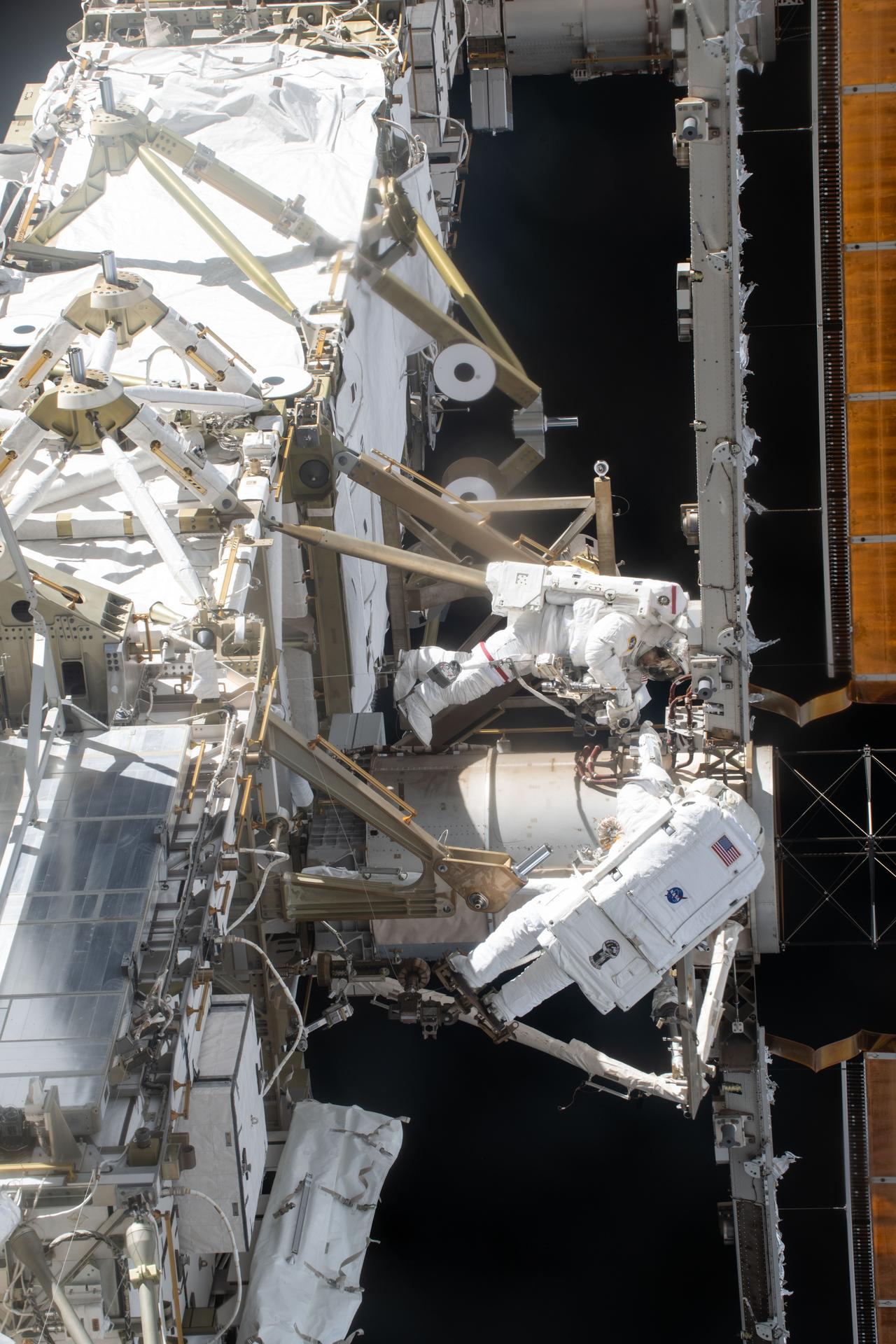
NASA astronauts Jessica Meir and Chris Williams, both Expedition 74 flight engineers, are pictured outside the International Space Station during a spacewalk to complete installation of a modification kit ahead of future solar array installation on the 2A power channel of the orbital outpost. It was William’s first spacewalk, Meir's fourth, and the 278th in support of space station maintenance, upgrades, and assembly.
NASA astronauts Jessica Meir (top) and Chris Williams are pictured during a spacewalk on March 18, 2026, to complete the installation of a solar array modification kit on the International Space Station.
NASA/Jack Hathaway

Spacewalk preparations filled the schedule aboard the International Space Station on Tuesday as a pair of astronauts gear up for next week’s external robotics repair job. CubeSat maintenance and eye checks rounded out the day for the Expedition 74 crew.

NASA flight engineers Chris Williams and Jessica Meir will once again wear their spacesuits and work together in the vacuum of space to repair a wrist joint on the Canadarm2 robotic arm starting at 7:35 a.m. EDT on Tuesday, June 30. The duo will spend about six hours and 40 minutes on the outside of the orbital lab replacing the malfunctioned wrist joint on Canadarm2 that has been in operation for over 25 years. NASA and the Canadian Space Agency (CSA) analyzed the issue and determined a spacewalk was necessary to replace the wrist joint with a spare already on the station. NASA and CSA officials will preview the upcoming spacewalk tasks during a news conference on NASA’s YouTube channel at 2 p.m. on Thursday, June 25.

Williams tried on his spacesuit Tuesday and tested the suit in its powered configuration with assistance from flight engineer Sophie Adenot from ESA (European Space Agency). The duo worked inside the Quest airlock checking the pressurized suit’s comfort and mobility and its communications and life support systems while engineers on the ground monitored.

Williams then joined Meir and reviewed on a computer a 3D interactive animation of the procedures and maneuvers they will use to return the Canadarm2 to full motion. Afterward, the pair took turns servicing emergency jet packs that will be installed on the spacesuits the day the spacewalk begins. Meir also installed batteries inside the pistol grip tools — designed especially for microgravity — they will use to work on the robotic arm.

Adenot and NASA flight engineer Jack Hathaway, who will monitor the spacewalkers from inside the orbital outpost, studied their spacewalk support roles including carefully adjusting the Canadarm2 into position during the repair work. Hathaway also had time for CubeSat work first removing a small satellite orbital deployer that was stowed inside the Kibo laboratory module’s airlock. Next, he installed a set of CubeSats inside the NanoRacks CubeSat deployer that will soon be placed outside Kibo’s airlock for deployment into Earth orbit. The tiny satellites were designed by university students for a variety of communications and technology investigations.

Station commander Sergey Kud-Sverchkov joined flight engineer Sergei Mikaev in the Harmony module for eye checks using standard medical imaging hardware. Doctors on the ground were examining how living in space affects the retina, lens, and cornea. Roscosmos flight engineer Andrey Fedyaev continued testing artificial intelligence tools to boost crew efficiency and communications in space.

Learn more about station activities by following the space station blog, @space_stationon X, as well as the ISS Facebook and ISS Instagram accounts.

Get the latest from NASA delivered every week. Subscribe here.

The post NASA Announces Spacewalkers for Robotic Arm Repair Work appeared first on NASA Science.

Former NOAA Employees Revive Climate.gov Web Site

23 June 2026 at 17:26
The database of federal global warming research recreates a website that was closed amid the administration’s broad retreat from climate science.

© Jason Andrew for The New York Times

The National Oceanic and Atmospheric Administration headquarters in Silver Spring, Md. The new site is an effort to preserve NOAA work previously available at climate.gov.

Brewing sustainable power from used coffee grounds

22 June 2026 at 12:00

What if your daily coffee routine could help power the world around you? 

Scientists are exploring ways to transform everyday waste, like spent coffee grounds, into materials for greener energy production. One promising method, called triboelectrification, uses electricity generated from motion. With this approach, materials that gain positive and negative charges are rubbed together to generate an electrical charge that can be harnessed by devices called triboelectric nanogenerators, or TENGs. TENGs are a greener alternative to fossil fuels because they generate electricity from everyday movement, such as pedestrian or vehicle motion. 

However, many negatively charged materials used in these devices are made from nonbiodegradable plastics, like PTFE, that persist as long-term waste in the environment. Over time, TENGs are also easily damaged by environmental conditions outside the laboratory. To create sustainable TENGs for real-world applications, scientists need to develop biodegradable and durable designs. 

To address this problem, researchers from Guangxi University in China developed a TENG that uses coffee grounds as its negatively charged material. Their design repurposes coffee waste to create a robust, low-cost device that can harvest energy.

To create the coffee-based material, the researchers mixed coffee grounds with a biodegradable glue-like plastic called polycaprolactone (PCL). They heated this mixture to 80°C (176°F) and molded it into cylindrical pellets. Then, they used a biodegradable plastic called polylactic acid (PLA) to 3D-print a fully enclosed box-like TENG containing vertical channels arranged like the parallel slots of a file organizer to house the pellets. 

Their TENG worked by shaking the coffee ground pellets within the channels, causing them to repeatedly rub against the PLA surfaces. Because coffee grounds strongly attract electrons, the pellets become negatively charged while the PLA surfaces become positively charged. This repeated pellet movement drove the electrons back and forth, generating electricity that was harvested through the TENG’s external circuit. 

To evaluate their TENG’s electrical performance, the researchers used an instrument that measures small amounts of electricity, called an electrometer. The electrometer measures electrical output based on how strongly the device pushes electrons through a circuit, called its voltage, how quickly electrons flow through it, called its current, and how many electrons flow through it, called its transferred charge.

The researchers verified that coffee ground pellets could serve as a viable TENG material by comparing their performance with commercial plastic pellets. The coffee ground pellet system produced a voltage of 0.6 Volts (V), a current of 18 nanoamperes (nA), and a charge transfer of 0.25 nanocoulombs (nC). This output was more than twice that of pure PCL pellets and over half that of nonbiodegradable PTFE. The researchers suggested that the negatively charged components of the coffee grounds altered the surface of the PCL to increase its charge generation and transfer. 

To optimize the TENG, the researchers experimented with different coffee grounds to PCL mass ratios, pellet lengths, and device vibrational frequencies. They found that their device achieved the best electrical performance at a 3:1 ratio of coffee grounds to PCL, with 8 millimeter long pellets, at a vibrational frequency of 2.6 Hertz. These conditions boosted the device’s electrical output to 4.7 V, 75 nA, and 1.3 nC, high enough to power tiny environmental sensors. 

The team also tested their device’s long-term durability by comparing its electrical performance before and after 6 months of storage. They observed only a 6% drop in voltage after storage, demonstrating that it could maintain stable long-term electrical performance for at least 6 months. 

Finally, to explore real-world applications, the researchers integrated 4 TENGs into a wind-energy harvesting system designed for remote island settings. The system used a wheel of rotating cups to capture wind energy and a sliding mechanism to convert it into repeated shaking of the TENGs. As the TENGs shook, the moving pellets inside generated enough electricity to power an LED light and a small weather-monitoring device. 

The researchers concluded that spent coffee grounds can function as biodegradable TENG materials while reducing dependence on fossil fuel-derived materials. By combining coffee waste with biodegradable plastics, the researchers created a TENG with good electrical performance, sustainability, and long-term stability. They suggested that with further development, the device could someday power road warning lights and provide sound-based alerts in remote areas.

The post Brewing sustainable power from used coffee grounds appeared first on Sciworthy.

❌