❌

Normal view

Lewis and Clark’s mercury-laced poop helped archaeologists trace their expedition

16 September 2026 at 13:58

The Corps of Discovery Expedition set out in 1804, directed by President Thomas Jefferson to survey the United States’ newly acquired Louisiana Purchase. The exploration forms a core piece of early U.S. history. Picture a scene from the journey: It’s a cool morning, the silence punctuated by birdsong and the rustle of the breeze through the cottonwood trees. To one side, Commander William Clark, his rifle leaning against a plum tree and his rucksack nearby, squats over a latrine pit, unceremoniously heeding the call of nature. 

The Louisiana Purchase constituted a territory of around 828,000 square miles, which the U.S. had bought from France in 1803. This was, it is worth noting, largely without the consent of the Indigenous groups who had lived there for millennia. The purpose of the expedition was threefold: to find a practical trade route westward to the Pacific coast, to explore and document the territory, and to stake a claim to the area before any other European powers butted in. Led by Meriwether Lewis and William Clark, the 40-man team, plus their guide Sacagawea, spent more than two years trekking westward.

Believe it or not, the holes that were used as toilets along the march to the Pacific have been invaluable for archaeologists and historians. This is thanks especially to a type of treatment included in the expedition’s medical kit. 

How Lewis and Clark went to the bathroom

Everybody poops—it’s a universal human need. But most accounts of the Corps of Discovery’s journey leave out the details of how the Corps members dealt with bathroom business. 

Each of the campsites where the expedition spent time had an accompanying latrine pit, situated well away from camp for sanitary (and privacy) reasons. The latrines would have seen a lot of use, because the Corps of Discovery’s diet wasn’t the best for gut health. 

Painting Lewis and Clark on the Lower Columbia (height: 24 in (60.9 cm); width: 28 in (71.1 cm)) by Charles Marion Russell; Opaque and transparent watercolor over graphite underdrawing on paper
American artist Charles Marion Russell painted this piece entitled “Lewis and Clark on the Lower Columbia” in 1905. Image: Public Domain

While the expedition members relied heavily on foraged and hunted food, they also carried backup rations. These included nearly 14,000 pounds of dry goods, including flour, salt, coffee, dried pork, and lard. All told, each member of the Corps ate around nine pounds of meat per day. All of that protein without lots and lots of fiber added up to frequent constipation. The untreated water they drank from rivers and creeks they encountered probably didn’t help their circumstances. Enter…Dr. Rush’s Bilious Pills. 

The mercury constipation pills known as “thunderclappers”

In 1803, as preparation for the upcoming expedition, William Clark traveled to Philadelphia for a two-week medical crash course with Dr. Benjamin Rush. Rush was a prominent physician in his day, and a Founding Father of the United States. His signature is one of the 56 on the Declaration of Independence.

Rush taught Clark some basic field skills, such as bone-setting and wound treatment. Clark left Philadelphia with new skills and a well-stocked medical kit that included 50 dozen of Doctor Rush’s Bilious Pills. These were compounded tablets about the size of a raisin, composed of powerful laxative ingredients. The pills were nicknamed “thunderclappers” because of the speed with which they took effect. The main ingredient of the thunderclappers was calomel, or mercury chloride. 

Mercury, historically also called quicksilver, has been thought to have curative effects for centuries. It’s a metal that is liquid at room temperature, which seems miraculous in and of itself. 

It was also a popular treatment for syphilis. This is not to say it was an effective treatment for syphilis. When it was administered for that disease, slowly in low doses, patients were more likely to die from mercury poisoning than from the STI. 

Drug jar for mercury pills. Italy, 18th Century. Polychrome Ointment jar, decorated with ruins and figures, for mercury pills. Italy, 1731-70.
This drug jar for mercury pills was made in Italy sometime between 1731 and 1770, several decades before Meriwether Lewis and William Clark set out on their historic expedition. Image: Science & Society Picture Library / Contributor / Getty Images Jaron Chubb

When taken in larger doses, though, as in the thunderclapper pills, it prompted a quick evacuation, the body’s way of ejecting poison. The men of the Lewis and Clark expedition used these pills freely as a way to compensate for their high-protein and low-fiber diet. As a result, all the latrine pits they used between the Midwest and the Pacific coast were laced with high concentrations of mercury. 

Lewis and Clark pooped mercury. Today archaeologists use it to map their route.

As many an environmentalist will caution, mercury does not decompose when it’s in the ground. It stays put, and is detectable through modern soil testing techniques. The 600-plus latrines dug and used by the Corps of Discovery form a trail of their own, marked by mercury. 

The expedition was a government-sanctioned military expedition, so Lewis and Clark both kept copious notes on every aspect of their travels. Historians have tried to pinpoint the locations of these camps based on the expedition journals, with minimal success. 

The only campsite that archaeologists have been able to confirm so far is the Traveler’s Rest camp, just outside of Missoula, Montana. This is all thanks to the remains of Dr. Rush’s Bilious Pills, which left enough mercury in the ground to differentiate the remains of a latrine pit (or “sink” as they were called in the 1800s) from animal burrows or agricultural ditches. 

Archaeology is all about reconstructing the lives of people in the past, and it’s worth remembering that this means all aspects of life. Our own bodily functions remind us that we all share complex and interwoven histories, and that even the most mundane parts of our lives can be important in the future. 

In That Time When, Popular Science tells the weirdest, surprising, and little-known stories that shaped science, engineering, and innovation.

Related 'That Time When' Stories

The post Lewis and Clark’s mercury-laced poop helped archaeologists trace their expedition appeared first on Popular Science.

Spectators Horrified as Hyrox Athlete Recreates “Two Girls One Cup” at Live Competition

15 September 2026 at 21:12

Conventional gym etiquette involves carefully wiping down the surfaces of any machines or equipment after using it. Businesses usually provide customers with wet wipes, or simple sprays and towels, to keep faux leather padding and other public surfaces pristine and sanitary.

But during professional events, that level of cleanliness isn’t always guaranteed. Organizers of a recent Hyrox competition in China, the spiritual successor of the Crossfit craze, have been accused of allowing participants be exposed to a trail of human waste — left behind, memorably, by the athlete who went on to win the competition.

As The Guardian reports, Australian world record holder Joanna Wietrzyk used shared stations, including rowing machines, sleds, and medicine balls, while “faecal matter” was “visibly running down her legs,” a horrifying incident vaguely reminiscent of one of the most infamous and reviled videos in internet history.

The incident drew an outburst of disgust across social media.

“I guess I’m never doing a Hyrox event,” one user wrote in a post on Bluesky. “Oh my god.”

“Saw a big Hyrox poster at the gym and all I thought of was that lady shitting herself,” another tweeted. “That’s their brand now.”

“This should result in a hard stop for the competitor because the public health risk is more important than the medal,” one user argued. “Hyrox has way too many shared touch points and surfaces to let this continue.”

Others used the opportunity to poke fun.

“A s****y situation for all parties and tough for Hyrox to save faeces,” another joked.

Organizers were criticized for allowing the competition to continue despite having clear bio-contaminant rules in place.

“Protocols designed for elite competition and mass participation became blurred, creating ambiguity in how they were applied and understood,” Hyrox World wrote in an statement, in one of the more acrobatic euphemisms we can recall.

Hyrox cofounder Moritz Fürste issued an apology on Instagram as well.

“Hyrox made a mistake by not reacting immediately during the race,” he wrote. “In the end it is my job to foresee these potential incidents — and I did not.”

“Of course, we started improving event processes,” Fürste went on, “plus making rulebook changes immediately.”

The shocking incident highlights the fact that professional endurance athletes soiling themselves isn’t as rare as it may seem. Long distance runners, for instance, are known to suffer from “runner’s diarrhea,” like Paula Radcliffe, who had to relieve herself in full view of the public during the 2005 London marathon.

Radcliffe still won with a time of 2:17:42 — albeit with a little less dignity than she started.

More on athletes: They Held a New Olympics Where Athletes Can Take as Many Drugs and Steroids as They Want, and the Funniest Possible Thing Happened

The post Spectators Horrified as Hyrox Athlete Recreates “Two Girls One Cup” at Live Competition appeared first on Futurism.

It’s almost time to airdrop rabies vaccines again

15 September 2026 at 17:34

Despite major strides in education and conservation efforts, rabies remains a serious public health issue. The Center for Disease Control (CDC) estimates around 90 percent of cases occur in wildlife, but their consequences still reverberate across ecosystems and routinely endanger humans. Combating the viral disease costs the United States over $300 million every year, but the hefty price tag is justified. Contracting one of the oldest known diseases invariably causes severe symptoms like abnormal sensations, violent movements, hydrophobia, and loss of consciousness. Rabies is fatal in virtually every documented case, and still kills around 59,000 people around the world each year.

Once primarily associated with dogs, rabies has since spread to wildlife such as raccoons, skunks, bats, and foxes—making the issue much more difficult to track and control. Since 1995, the United States Department of Agriculture (USDA) has overseen a National Rabies Management Program in cooperation with state and federal departments, university researchers, and other organizations. After over 30 years of work, one of the most preferred mitigation methods is regularly placing oral rabies vaccine (ORV) bait in strategically selected wildlife areas. The easiest way to spread millions of bait blocks across thousands of square miles? Airdrops.

Blue gloved hand holding raccoon rabies vaccine bait packets
The bait isn’t toxic, but the USDA recommends using gloves to handle it. Credit: USDA

Next month, the USDA will once again begin tossing fishmeal-coated ORV packets from planes throughout the Southeastern states of Virginia, North Carolina, Tennessee, Georgia, and Alabama. These tiny edibles are specifically crafted to target the most prominent rabies variants seen in raccoons, coyotes, and gray foxes.

“The goal is to prevent westward spread of raccoon rabies by creating a continuous vaccine barrier that stays in front of rabies occurrence on the landscape,” a USDA spokesperson recently told FOX Carolina News. The representative added that although the distribution’s scope doesn’t currently include South Carolina, they are “very close to establishing an ORV area in South Carolina counties bordering North Carolina and Georgia in the next few years.”

If all goes as planned, the National Rabies Management Program’s geographic scope will shrink over time as the disease is regionally eliminated. Until then, anyone who accidentally comes into contact with an ORV bait brick shouldn’t worry about contracting the disease. The vaccine inside each brick is proven safe for over 60 animal species, including cats and dogs. If you happen to locate one in a residential area, the USDA recommends to either simply leave it alone, or use gloves to move it to a more remote location for its intended recipients. Always wash your hands thoroughly after handling ORV bait—and try to ignore the pungent odor of fishmeal.

The post It’s almost time to airdrop rabies vaccines again appeared first on Popular Science.

Laptop Toasts Boy

13 September 2026 at 16:02

Here’s a hazard you probably didn’t expect from too much screen time.

Two doctors in the UK reported in the journal BMJ Case Reports the bizarre case of a young boy whose laptop literally toasted his skin over weeks and maybe even months of using it. Thankfully, the kid’s fine and was never in any real pain, so we can dive into this medical oddity without feeling too guilty about it.

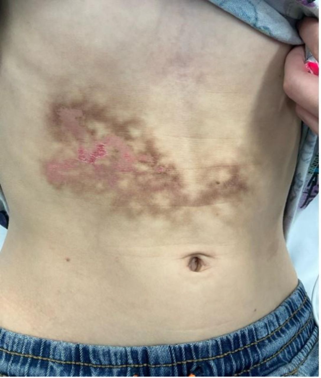
According to the eyebrow-raising paper, spotted by Ars Technica, the boy was first brought into an emergency department with reddish markings over his abdomen in a lace-like pattern. Nurses first described it as “spontaneous bruising,” but they didn’t find skin abnormalities anywhere else on his body. The boy also wasn’t in any pain, and didn’t have any history of trauma or illnesses that could explain the markings.

When doctors did a chest x-ray, they found some kind of build up in his lung, though it was unclear what it signified. The boy admitted that he got pushed into a chair while play-fighting with his siblings a week ago, leading the doctors to search for signs of a traumatic injury. But after a CT scan, they didn’t find any. They weren’t completely sure of what was going on, but the doctors discharged him with antibiotics for a suspected lung infection.

The boy was back several weeks later, though — and the rash was even worse. Parts of his abdomen were pinkish, exhibiting clear signs of skin breakdown, the doctors wrote, with mottled patches of dark brown. (You can view a photo, at your own peril, here, via Ars.)

That’s when the doctors began to suspect some kind of heat damage. But when they asked the boy about heat sources, nothing turned up. 

Then, as they continued to question him, the boy mentioned something crucial: he was home-schooled and did most of his assignments on his laptop, sometimes for eight hours a day, often while charging. And of course, he usually rested the laptop on his torso while lying down.

This supported a diagnosis of a erythema ab igne (EAI), also known as “toasted skin syndrome.” It’s caused by repeated, close exposure to low heat, which slowly damages the skin over time. Patients often get it from heat pads and space heaters, but a warm laptop can cause it, too. It causes a distinctly lace-like rash, which explained the boy’s symptoms.

EAI is benign, and in most cases, its rashes disappear on their own once exposure to a heat source is stopped, taking weeks to months to heal. Once properly diagnosed, the boy’s rash was dressed, and he came back to the hospital several months later to confirm that he had fully healed. Now that he’s okay, he might try studying at a desk instead.

More on medical oddities: Man Announces That He Has Synthesized a New Schizophrenia Treatment in His Garage, Based on a Formula Devised by ChatGPT

The post Laptop Toasts Boy appeared first on Futurism.

America’s Health Care System Is So Screwed That Patients Are Flying to China for Cancer Treatment

13 September 2026 at 15:01

The myriad horrors of the US healthcare system have spawned generations of medical tourism from the wealthiest country on Earth to the periphery: Cuba, Mexico, Costa Rica, Canada, Turkey, and Thailand, to name just a few.

Now, Shanghai is quickly rising as one of the top destinations for cancer patients, as China’s biotech sector has spun an experimental treatment developed in the US into a standalone industry.

New reporting by the Wall Street Journal took a look at this fast-growth industry, centered around chimeric antigen receptor T-cell therapy, or CAR-T for short.

CAR-T is essentially a personalized form of immunotherapy, in which a patient’s own white blood cells are engineered to identify and attack specific proteins found in cancer cells. Though the process was first developed by US biotech giants, their inability — or unwillingness — to lower the cost and increase supply of the treatment has led to shortages throughout the country.

That’s where China comes in. As director of the CAR-T program at the University of Texas MD Anderson Cancer Center in Houston Sairah Ahmed told the WSJ, this therapy can cost around $150,000 to $230,000 in China — still a life-altering bill, but a sizeable discount compared to an insurmountable $550,000 to $850,000 in the US.

This, Ahmed says, is why she advises some patients to seek out treatment in the People’s Republic.

“China has basically done to CAR-T cell therapy the same thing that it’s done to other industries, which is, they’ve industrialized it,” Ahmed explained.

China’s CAR-T services aren’t just cheaper, either. They’re also faster, and come with a host of complimentary after-care treatments, the WSJ notes. As MD Anderson Cancer Center’s explainer on CAR-T therapy advises, the process of extracting and bio-engineering a patient’s cells to fight cancer can take over three weeks in the US. Compare that to China, where the WSJ reports labs near Shanghai can “crank out” the therapy in just a matter of days.

An important caveat here is that CAR-T cell therapy isn’t a catch-all for every type of cancer, and it’s typically only administered after other treatments have failed. It’s not exactly a walk in the park, either, requiring months of recovery and check-ups from a range of physical, neurological, and psychological health specialists. Still, if you had to go through with it, your first step should clearly be pricing out some flights to China.

More on healthcare: Bougie Health Startups Offering Full-Body Diagnostic Scans for Rich People While the Rest of Us Suffer a Crumbling Healthcare System

The post America’s Health Care System Is So Screwed That Patients Are Flying to China for Cancer Treatment appeared first on Futurism.

Bizarre New Websites Are Designed Specifically to Spark Dopamine in Your Brain

13 September 2026 at 13:02

As far as completely pointless ways to kill time on your phone, this is certainly one of the weirdest on record.

Behold the all-new invention of so-called “dopamine sites” — digital domains where you can browse and hunt for deals on nonexistent wares and services. Clothes, food delivery, you name it: you can pretend-shop for it all. 

You don’t actually buy anything, crucially. You’re just pretending to have money to buy the things you want. It’s window-shopping reimagined for the age of doomscrolling.

But it’s also a little more than that. These cleverly-built sites simulate the experience down to checking out and “tracking” your shipment or delivery, and their absurdly realistic rituals can help people become more conscious of their spending habits. Supposedly. 

The trend, according to the New York Times, originated in South Korea this spring, when Park Seohyun, a 21-year-old mechanical engineering student in Seoul, created a fake food-delivery site called “Food Never Arrives.” (It’s now an app).

Originally intended as a joke, she says, it lets you not only order nonexistent food, but provides you an estimate of the money and calories you saved by skipping on the real thing. 

In a wintry economic climate, it became an unexpected hit.

“People began sharing it online and discussing whether it actually helped them resist ordering food,” Park told the NYT.

A wave of imitators followed. This summer marked the launch of “FakeHaul,” a dopamine site built by a Microsoft engineer that convincingly imitates shopping on Temu. Two developers in India inspired by Park’s site, Purvansh Parmar and Anshul Sharma, created “Food Never Comes,” which simulates food deliveries but provides real recipes for the food you didn’t actually order. Parmar and Sharma told the newspaper that their site had more than 2.7 million views since launching in June.

Whether these dopamine sites provide anything more than fleeting, empty pleasures is something experts are more skeptical about. Ravi Dhar, director of the Yale Center for Customer Insights, told the NYT that the “element of looking forward brings a lot of the pleasure, and we often crash after an event or feel let down.” Lingguo Xu, a behavioral economist studying anticipatory utility, warned that the dopamine tricks could quickly get old. And by simulating consumption, you risk developing “stronger cravings.”

Some take the original idea beyond getting high on simulated shopping. Ko Seojin, another South Korean developer, created a website for taking virtual smoke breaks called Damta World. You click to start smoking an animated cigarette, which starts burning on screen. You can “whisper” disappearing messages to other smokers on break, while a counter keeps track of the number of cigarette butts that have been tossed on the office rooftop each day.

Ko told the NYT that 150,000 people visit each month, and she says that some users have become real-life friends after meeting on the website. Those friendships, we hope, are something that’ll last longer than any fake-shopping dopamine hit.

More on your brain on tech: Psychologist Says AI Is Causing Never-Before-Seen Types of Mental Disorder

The post Bizarre New Websites Are Designed Specifically to Spark Dopamine in Your Brain appeared first on Futurism.

GLP-1 Users Are Being Cruelly Shamed by Their Own Families

12 September 2026 at 17:01

GLP-1 weight loss drugs are changing lives in more ways than one.

In a new piece for The Guardian, the newspaper’s readers share how the weight loss journey that drugs like Wegovy, Ozempic, and Mounjaro have set them down on have drastically affected their interpersonal relationships — sometimes for the better, and sometimes for the worse. 

Many of the GLP-1 users seem to harbor a sense of shame, hiding their use of the drugs. In their minds — and perhaps in the minds of those around them — they undermine the sense of accomplishment that comes with losing weight the old fashioned way, instead feeling that they “cheated” or are “being dishonest” about how they achieved their goals. 

One parent wrote of how their 20-year-old daughter felt “worried, angry, and jealous” after she discovered they were hiding their use of Wegovy from her. “We had discussed weight, identity and GLP-1s, but I was so ashamed of going on it that I didn’t want to tell anyone,” they wrote, noting how food was a “huge part of our family life.” They wondered to themselves: “What kind of signal am I giving her and her brothers?” 

Though they feel “mostly positive” about losing nearly thirty pounds using the drug, they added, they also can’t shake the feeling that they’re “cheating.”

“So much of my insecurity about my weight and appearance is offloaded on to an injectable pen,” they added.

Another wrote that despite their friends feeling pleased about their weight loss, they’ve kept their GLP-1 drugs secret for nearly two years. Their partner, they believe, “has felt insecure at times because of my glow-up.”

“I don’t want people to think I’ve cheated. I guess there is still fat shame somewhere,” they wrote.

Others don’t feel this guilt, but are frustrated that their spouses don’t realize how transformative the drugs have been for them. One complained that their husband was so unsupportive of their use of GLP-1s that they no longer could share their experience with him.

“This has been a life-changing drug so far, and I can’t share it with the person I’m closest with,” they lamented. 

Though they’ve struggled with obesity their whole life, their “husband scoffs at characterizing obesity as a chronic and recurring disease” and “thinks people should just make better lifestyle choices and stop making excuses — which tells me a lot about how he views me, too.”

These downsides aren’t unique to GLP-1s. Significant weight loss has long been linked to divorce, and that doesn’t necessarily mean that someone who’s slimmed down is now feeling their oats. Weight loss can come with dramatic changes to a person’s lifestyle — from the food they eat and the exercise they do to how they socialize — and that can be a lot for someone close to them to adjust to.

“Relationships tend to develop a sense of equilibrium and predictability over time,” Lindsey Berisha, a clinical psychologist who’s worked with patients using GLP-1s for over a decade, told The Guardian. “When one partner undergoes a significant change, it can shift that equilibrium, and even positive change can create uncertainty for both partners.”

GLP-1s, of course, have made achieving this kind of significant weight loss much more accessible, which is why we’re now starting to hear about “Ozempic divorces.”

More on GLP-1s: GLP-1 Drugs Appear to Prevent Cancer, New Research Finds

The post GLP-1 Users Are Being Cruelly Shamed by Their Own Families appeared first on Futurism.

Five-Year Disease Progression in Synuclein-Positive Sporadic Parkinson’s Disease

11 September 2026 at 12:06

Parkinson’s disease has long been a diagnosis of observation and inference. Doctors watched for tremor, rigidity, and slowness of movement, and only at autopsy could the telltale clumps of alpha-synuclein protein—the pathological signature of the disease—be confirmed inside the brain. That diagnostic fog may finally be lifting. A new five-year study drawing on the landmark Parkinson’s Progression Markers Initiative (PPMI) has followed patients whose disease was defined not by their symptoms alone, but by biology: a positive cerebrospinal fluid seed amplification assay, the test that detects misfolded alpha-synuclein circulating in the nervous system. The results, published in Annals of Clinical and Translational Neurology, offer one of the clearest longitudinal pictures yet of how biologically confirmed Parkinson’s disease actually progresses in the era of modern treatment—and the findings are already reshaping how scientists think about staging, enrollment in clinical trials, and the very definition of the disease.

The research team set out with two central questions. First, what happens clinically to patients who test positive for synuclein seeding in their spinal fluid over five years of careful observation? Second, does a patient’s baseline stage on a new biological staging system—the Neuronal Synuclein Disease Integrated Staging System, or NSD-ISS—predict how quickly they will cross meaningful clinical milestones? Both questions cut to the heart of a quiet revolution in neurology. For more than a century, Parkinson’s was classified by what patients looked like in the examination room. Now, thanks to validated biomarkers, researchers can classify it by what is happening at the molecular level, potentially years before disabling symptoms emerge.

The seed amplification assay, or SAA, is the technological engine behind this shift. The test exploits a peculiar property of misfolded alpha-synuclein: it acts as a template that recruits normal, healthy synuclein proteins and forces them to misfold as well, seeding the aggregates known as Lewy bodies that riddle the brains of Parkinson’s patients. In the laboratory, a tiny sample of cerebrospinal fluid is mixed with synthetic alpha-synuclein and monitored for hours. If pathological seeds are present, the reaction accelerates into a detectable fluorescence signal. The assay has been validated across multiple international cohorts and, critically, against postmortem brain tissue, giving neurologists a window into pathology they previously could only glimpse after death. A complementary technique—detecting phosphorylated alpha-synuclein in small skin biopsies—has added a second, less invasive line of biological evidence.

Armed with these tools, two research groups have proposed frameworks for redefining Parkinson’s disease biologically. The SynNeurGe criteria classify patients by the combined presence of pathological alpha-synuclein biomarkers, neuroimaging evidence of neurodegeneration, and disease-relevant genetic variants. The Neuronal Synuclein Disease criteria take a parallel approach, defining disease by the presence of pathological synuclein as measured by a validated biomarker, with or without evidence of dopaminergic dysfunction detected through dopamine transporter imaging. The integrated staging system then arranges these biological anchors along a seven-stage ladder: Stage 0 reserved for carriers of fully penetrant mutations in the SNCA gene; Stages 1A and 1B for people with synuclein pathology but no symptoms, depending on whether dopaminergic dysfunction is present; Stages 2A and 2B for those with subtle signs that stop short of functional impairment; and Stages 3 through 6 capturing progressively severe clinical disability.

PPMI, the international observational study launched in 2010, provided the ideal laboratory for testing whether this staging framework means anything in the real world. The researchers focused on the sporadic Parkinson’s cohort: participants diagnosed within two years of enrollment who had never taken dopaminergic medication, whose examinations showed cardinal motor features, and whose dopamine transporter scans confirmed the characteristic deficit in the striatum. Crucially, the team selected only those participants who met biological NSD criteria through a positive CSF seed amplification assay, recruited before 2020 to guarantee at least five years of follow-up. This design deliberately stripped away a longstanding source of noise in Parkinson’s research: the clinical heterogeneity that arises when a “Parkinson’s” diagnosis might actually encompass unrelated neurodegenerative processes that mimic the disease but follow entirely different biological courses.

Over the five-year observation window, participants underwent an unusually thorough annual workup. Motor and non-motor function was tracked with the Movement Disorders Society Unified Parkinson’s Disease Rating Scale across all four of its parts, alongside the Hoehn and Yahr staging scale and the Schwab and England activities of daily living score. Smell was measured with the University of Pennsylvania Smell Identification Test, autonomic function with the SCOPA-AUT, mood with the Geriatric Depression Scale, and REM sleep behavior disorder risk with a dedicated screening questionnaire. Cognition was assessed with the Montreal Cognitive Assessment and, from the study’s third year onward, formal clinician diagnoses of normal cognition, mild cognitive impairment, or dementia. Medication burden was quantified as levodopa equivalent daily dose, and dopamine transporter imaging was repeated at years one, two, and four, quantified both in the putamen—the region most affected in Parkinson’s—and across the striatum as a whole.

The broad message from the five-year trajectories is one of measurable, biologically anchored progression. Patients recruited as freshly diagnosed, biologically confirmed sporadic Parkinson’s patients showed the expected decline across motor scales and dopaminergic imaging, with the earlier PPMI analysis by Simuni and colleagues having already documented significant—though modest—correlation between worsening clinical scores and falling DAT binding over five years. What the new analysis adds is the biological filter: by restricting the cohort to synuclein-seeding-positive individuals, the study reduces the contamination from look-alike conditions that has historically muddied progression estimates. When a cohort is defined by its underlying pathology rather than its outward symptoms, the resulting disease course becomes a truer reflection of what alpha-synuclein itself does to the nervous system over time.

Perhaps the most consequential findings concern prediction. If the NSD-ISS staging system is to earn its place in research clinics and, eventually, in therapeutic trials, it must do more than organize patients neatly on a page—it must forecast what comes next. The study analyzed whether a patient’s baseline stage predicted survival and the time required to reach clinically meaningful disease milestones: crossing thresholds on the clinical rating scales, advancing in Hoehn and Yahr stage, slipping in daily living independence, or developing cognitive impairment. The logic is straightforward and powerful. A patient sitting at Stage 2B—with confirmed synuclein pathology, dopaminergic dysfunction, and subtle signs but no functional impairment—should, in theory, march down the staging ladder at a predictable pace. Demonstrating that baseline stage genuinely stratifies risk would give trial designers a rational tool for enrichment, allowing them to recruit patients at the stage where a candidate drug is most likely to show benefit.

That trial-design implication is not academic. Across neurodegenerative disease research, therapeutic development is pivoting decisively toward biomarker-defined enrollment. The bitter lessons of Alzheimer’s trials—where anti-amyloid therapies only proved effective once trials recruited based on biological confirmation rather than syndrome alone—have not been lost on the Parkinson’s community. Drugs targeting alpha-synuclein directly, whether through immunotherapy, aggregation inhibition, or other mechanisms, are entering trials that increasingly require positive seed amplification assays or other biological confirmation as a gate for entry. A validated staging system that predicts five-year trajectory would allow sponsors to select participants early enough in the disease process for neuroprotective strategies to matter, while reserving later-stage patients for symptomatic interventions. The five-year PPMI data provide exactly the kind of naturalistic benchmark that such enrichment strategies demand.

The study also marks a conceptual milestone: the description of Parkinson’s disease under contemporary management. Patients diagnosed today are treated differently than those diagnosed twenty years ago, and their disease course may differ as a result. Describing outcomes in a biologically defined, prospectively observed cohort establishes a modern baseline against which future disease-modifying therapies can be judged. When an experimental drug claims to slow progression, the comparison will be against trajectories like those documented here—precise, biomarker-anchored, and free of the diagnostic uncertainty that plagued earlier natural history studies. In that sense, the paper functions simultaneously as a clinical report and as a foundation stone for the next generation of Parkinson’s trials.

What emerges from five years of watching synuclein-positive patients is a disease that can now be seen, staged, and tracked before it fully announces itself. The combination of CSF seed amplification assays, dopamine transporter imaging, and structured clinical assessment has converted a syndrome defined in the examination room into a biological disease measurable in the laboratory. If the staging system validated in this cohort continues to predict who declines fastest and who reaches milestones soonest, neurologists may one day tell a newly diagnosed patient not only what they have, but with unprecedented confidence what lies ahead—and researchers may finally test neuroprotective drugs in the early biological window where they stand the best chance of changing the story. For a disease that has resisted precise definition since James Parkinson first described it in 1817, that is a transformation worth watching.

Subject of Research: People

Article Title: Five-Year Disease Progression in Synuclein Seeding Positive Sporadic Parkinson’s Disease

Article References: Gonzalez‐Latapi, P., Gochanour, C., Choi, S. H., Cho, H., Caspell‐Garcia, C., Coffey, C., Brumm, M., Lafontant, D.-E., Xiao, Y., Tropea, T., Seibyl, J., Tanner, C., Venuto, C. S., Kieburtz, K., Chahine, L. M., Poston, K. L., Siderowf, A., Marek, K., Simuni, T., & The Parkinson's Progression Markers Initiative (2026). Five‐Year Disease Progression in Synuclein Seeding Positive Sporadic Parkinson's Disease. Annals of Clinical and Translational Neurology, 13(9), 1791-1806. https://doi.org/10.1002/acn3.70323

Image Credits: AI Generated

DOI: 10.1002/acn3.70323

Keywords: Parkinson’s disease, alpha-synuclein, seed amplification assay, Neuronal Synuclein Disease, NSD-ISS staging, PPMI, biomarkers, dopamine transporter imaging, disease progression

Boy Developed 'Toasted Skin' Condition From Using a Laptop Every Day

By: BeauHD
11 September 2026 at 12:00
A medical report published earlier this week says a boy in the U.K. developed erythema ab igne, or "toasted skin syndrome," after resting a laptop on his abdomen for up to eight hours a day, often while it was charging. "The marks were described as being in a patch about 15 centimeters (six inches) wide, made of flat, reddish-brown 'interlacing lines forming irregular circles and a lace-like morphology,'" reports Ars Technica. From the report: Based on the appearance, the doctors started reconsidering some sort of bruising and started asking him more questions. This is when he mentioned that he was homeschooled and used a laptop for his schoolwork. He said he typically did his work by resting his laptop on his abdomen while he worked, sometimes for up to eight hours a day, and often while the laptop was charging. Then they understood what it was. The doctors diagnosed the boy with erythema ab igne (EAI), which translates from Latin as "redness from fire" and is also sometimes called "toasted skin syndrome." This is a skin condition caused by repeated, prolonged exposure to low-grade heat -- heat not hot enough to cause actual burns. It's most often seen in adults, and common causes include using heating pads or hot water bottles for chronic pain or sitting too close to space heaters. Some people also develop it from certain occupational hazards, like glassblowing and metal work. EAI produces a lace-like rash, just like the boy's. It's unclear how EAI develops exactly. But researchers hypothesize that chronic heat exposure leads to damage to superficial blood vessels and the release of red blood cells, forming the tell-tale red reticulated pattern. While the boy's condition looked ominous, the good news is that EAI is generally benign and reversible -- once you remove the heat source. For severe or long-term conditions, some pigmentation and scarring can remain. But the boy's parents checked back in over several months, reporting that the rash faded and then completely resolved. Hopefully, he also got some sort of laptop stand.

Read more of this story at Slashdot.

Neighborhood environment linked to frailty and PTSD in 9/11 responders

11 September 2026 at 11:11

Twenty-five years after the September 11 terrorist attacks, researchers at the Icahn School of Medicine at Mount Sinai have uncovered evidence that the environments in which World Trade Center responders live today may be quietly shaping their risk of frailty, post-traumatic stress disorder, and depression. The study, published in Nature Communications, is the first to examine how neighborhood-level environmental exposures later in life may interact with the chemical and psychological injuries sustained during the disaster response, adding a new dimension to one of the most intensively studied disaster health cohorts in the world.

The research team analyzed data from 18,734 responders who both live in New York City and receive care through one of the World Trade Center Health Program Clinical Centers of Excellence. These responders represent a population that experienced an extraordinary confluence of toxic exposures—dust, combustion products, airborne particulates, and a cocktail of known and suspected carcinogens—alongside severe psychological trauma. While decades of prior research have documented elevated rates of respiratory disease, cancer, and mental illness in this group, far less attention had been paid to whether the everyday conditions of their neighborhoods might compound or buffer those effects as responders age.

To answer that question, the researchers linked each responder’s residential address to annual estimates of three environmental factors: air pollution, high ambient temperatures, and neighborhood greenness, the last typically quantified through satellite-derived vegetation indices. This exposure-assessment approach, common in the field of environmental epidemiology, allowed the team to characterize the conditions each participant encountered where they lived year after year, rather than relying on broad regional averages. The researchers then examined whether those exposures were associated with the probability of being classified as frail, and with diagnosed PTSD and depression.

The results were striking in their consistency. Responders living in hotter neighborhoods—areas that experience more extreme heat days, a burden that falls disproportionately on densely built urban districts with impervious surfaces and little tree canopy—showed an elevated risk of frailty, a geriatric syndrome marked by diminished physiological reserve and heightened vulnerability to stressors. Elevated concentrations of air pollution were likewise associated with increased risks of both frailty and adverse mental health outcomes. Frailty itself is increasingly understood as a whole-body phenomenon, involving chronic inflammation, immune dysregulation, and cumulative cellular wear, all of which are plausible biological pathways through which heat and pollution could accelerate aging.

The mental health findings carried equal weight. Higher pollution and heat exposure were associated with increased odds of PTSD and depression, while responders living in greener surroundings—those with more parks, trees, and vegetated land near their homes—showed lower risks of all three conditions. This inverse association with greenness aligns with a substantial body of literature suggesting that access to green space can reduce psychological stress, encourage physical activity, dampen inflammation, and moderate urban heat, though the observational design of the study means that causation cannot be definitively established.

Perhaps the most provocative finding concerned interaction effects. The researchers found that responders who had experienced higher levels of World Trade Center–related chemical and psychological exposure appeared more vulnerable to the effects of environmental stressors in their current neighborhoods. In other words, the disaster and the decades that followed may not be independent chapters in these individuals’ health histories. Instead, responders already carrying the biological imprint of trauma and toxic dust—including altered inflammatory profiles, epigenetic changes, and persistent hypothalamic-pituitary-adrenal axis dysregulation—may enter later life with less reserve to absorb additional environmental insults. Everyday exposures that might be merely uncomfortable for a healthy middle-aged adult could, for these individuals, translate into measurable acceleration of physical decline and psychological deterioration.

“Twenty-five years after 9/11, we are still learning about the factors that influence the health of those who responded to the attacks,” said Helena Furman Krasnov, PhD, postdoctoral researcher at the Icahn School of Medicine and the study’s first author. “Our findings suggest that everyday exposures, including heat, air pollution, and access to green space, may contribute to responders’ long-term physical and psychological well-being. Understanding these factors could help identify new opportunities to support healthy aging in this population.”

The study’s senior and corresponding author, Maayan Yitshak-Sade, MPH, PhD, Associate Professor of Environmental Medicine at Mount Sinai and a member of the Institute for Exposomic Research, framed the work within the broader exposome paradigm—the effort to capture the totality of environmental exposures an individual accumulates over a lifetime. “Our findings suggest that the health consequences of 9/11 may not end with the disaster itself,” she said. “For responders already carrying the biological effects of trauma and toxic exposures, everyday environmental conditions may represent an additional source of stress on the body and mind. These findings highlight the importance of considering where people live, not just what they experienced during the disaster.”

That framing underscores a conceptual shift in disaster medicine. Traditional models tend to treat a catastrophic event as a discrete exposure with a defined aftermath. The Mount Sinai findings instead suggest that disaster-related health effects may unfold across an entire life course, modified by conditions that have no obvious connection to the original event. A responder’s risk of frailty in 2026 may reflect not only the hours spent at Ground Zero, but also the temperature of the summers they have endured since, the particulate matter drifting through their streets, and whether a leafy park sits within walking distance of their apartment.

The study arrives at a moment when urban environmental health has become a pressing policy concern. Extreme heat now kills more Americans annually than any other weather phenomenon, and cities including New York have launched ambitious initiatives to expand tree canopy, create cool corridors, and retrofit public housing with resilient cooling infrastructure. Air pollution, even at levels within regulatory limits, continues to be linked to cardiovascular, respiratory, and neurocognitive harm. If the associations identified in this cohort hold up under further scrutiny, they could strengthen the case for targeted environmental interventions—not as amenities, but as clinical supports for a vulnerable population.

The implications also extend well beyond the World Trade Center community. The researchers point out that survivors and responders from other disasters—wildfires that blanket entire regions in smoke and ash, industrial accidents that release toxic plumes, building collapses that bury rescue workers in dust—may face the same dual burden of physical and psychological exposures that continues to shape health long after the initial event. Climate change is intensifying heat waves and wildfire seasons simultaneously, meaning that disaster-affected populations may increasingly find themselves living in precisely the kinds of hostile urban microclimates the study identifies as risky. The framework developed here could inform how health surveillance programs for disaster cohorts are designed, prompting them to collect residential and environmental data alongside clinical outcomes.

The authors are careful to note the study’s observational nature. Associations between neighborhood environment and health can be confounded by socioeconomic status, housing quality, access to care, and the self-selection of healthier individuals into greener, quieter neighborhoods. The investigators controlled for available covariates, and the sheer size of the cohort lends statistical power, but randomized evidence on green space exposure is practically impossible to generate, and residual confounding remains an inherent limitation of the design. Longitudinal follow-up, and ideally natural-experiment analyses—such as the addition of new parks or major infrastructure changes—will be needed to firm up causal inference.

That work is already on the horizon. The team plans to investigate whether beneficial environmental factors, including green space and access to healthy food, can actively reduce the long-term health effects associated with World Trade Center–related exposures—a question that shifts the framing from risk identification to potential intervention. If confirmatory studies support the protective role of greenness, the findings could justify clinical guidance for responders, and potentially for other disaster-exposed populations, that goes beyond medication and monitoring to encompass the places where patients live.

For a population that has already given so much and borne so much, the idea that healing might partly depend on the landscape outside the front door is both sobering and hopeful. It suggests that some of the damage inflicted on September 11, 2001 may not be fixed, but modifiable—and that the next chapter of care for responders could be written not only in clinics, but in city planning offices, park commissions, and the slow, patient work of greening the neighborhoods where aging heroes live out their lives.

Subject of Research: People

Article Title: Urban Exposures, Frailty, and Mental Illness in World Trade Center Health Program Responders

Article References: Krasnov, H., Hung, W., Knobel, P., Kloog, I., Ko, F., Thompson, H., Colicino, E., Teitelbaum, S. L., Just, A. C., & Yitshak-Sade, M. (2026). Urban exposures, frailty, and mental illness in World Trade Center Health Program responders. Nature Communications, 17(1), Article 9449. https://doi.org/10.1038/s41467-026-77401-7

Image Credits: AI Generated

DOI: 10.1038/s41467-026-77401-7

Keywords: World Trade Center responders, 9/11 health, frailty, PTSD, depression, air pollution, extreme heat, green space, exposome, environmental health, Nature Communications, Mount Sinai

Cutting Two Amino Acids Slowed Prostate Cancer in Mice

10 September 2026 at 15:05
Illustration of Lipid Rich Prostate Cancer CellsA newly identified link between amino acid metabolism and cholesterol production may help prostate tumors adapt to hormone therapy. Prostate cancer can find ways around treatments designed to deprive tumors of the hormones they need to grow. Weill Cornell Medicine investigators have now traced one possible route of resistance to amino acid metabolism, where a [...]

New EAMA highlights showcase 30-year cohort generation research findings

10 September 2026 at 13:53

Thirty years after a small group of European professors of geriatric medicine first warned that the continent’s fastest-growing population was being cared for by physicians who had never been formally trained to care for them, the program they created to fix the problem is marking its anniversary with a scientific milestone of its own. A new special article published in European Geriatric Medicine documents the research output of the most recent graduating cohort of the European Academy for Medicine of Ageing, or EAMA, and in doing so offers a rare longitudinal measure of how a structured, pan-European leadership program in geriatric medicine translates into scholarly productivity, professional advancement and, ultimately, better care for older people.

The new paper, titled “‘Highlights of latest EAMA’: generation research outputs of the 30-year anniversary cohort,” was authored by Thomas Münzer of the Geriatric Clinic St. Gallen in Switzerland, Hannes B. Stähelin of Basel, and Barbara van Munster of the University of Groningen Medical School in the Netherlands, writing on behalf of the EAMA Board. It arrives at a moment when the demographic arithmetic of Europe has made the question of geriatric capacity more urgent than ever: the proportion of people over 65 continues to climb, multimorbidity, frailty and polypharmacy have become the daily reality of hospital medicine, and health systems across the continent still struggle to recruit and retain physicians trained specifically in the medicine of ageing. The article’s premise is simple but rarely examined with this degree of rigor — if you invest systematically in the education of mid-career geriatricians, what do you get back?

To understand the significance of the anniversary cohort’s research output, one has to go back to the early 1990s, when the group of European Professors in Medical Gerontology published a stark assessment in the journal Age and Ageing. Writing in 1994, Stähelin and colleagues documented that medical gerontology was taught unevenly, and in many countries not at all, in European medical schools. Geriatric medicine, despite being one of the few specialties whose patient population was guaranteed to expand every single year, was frequently a subordinate unit within internal medicine, staffed by physicians who had acquired their expertise on the job rather than through dedicated training. The professors’ conclusion was that Europe needed not merely more geriatricians but a new cadre of academic leaders — clinicians who could teach, publish, shape curricula and advocate for the specialty within their national health systems.

That diagnosis led, in 1995, to the founding of the European Academy for Medicine of Ageing, an intensive postgraduate program deliberately modeled on leadership academies rather than conventional continuing medical education. EAMA’s structure has remained remarkably stable across its three decades: a small, competitively selected cohort of physicians, typically already experienced clinicians, is brought together for a series of residential courses spread over roughly two years, interspersed with mentored projects that require participants to produce original scholarly work. The curriculum spans the full breadth of the medicine of ageing — from the biology of ageing and the physiology of frailty, through geriatric syndromes such as falls, delirium, incontinence and sarcopenia, to the organizational science of acute geriatric units, nursing home medicine, rehabilitation and end-of-life care. Crucially, faculty are drawn from across Europe, exposing participants to radically different national models of geriatric care and building a professional network that persists long after the formal courses end.

The “reloaded” reform of the program, described in a 2018 paper by Roller-Wirnsberger and colleagues in European Geriatric Medicine, formalized what three decades of experience had taught the academy’s faculty. That paper set out explicit competencies and standards for a European leadership program in geriatric medicine, defining what a graduate of EAMA should be able to do: not just diagnose and treat, but design services, lead multidisciplinary teams, mentor juniors, conduct and critique research, and represent the specialty at institutional and governmental levels. The competency framework aligned the academy with modern medical-education theory, in which learning objectives are specified in advance and assessed rather than assumed. It is against this framework that the anniversary cohort’s performance, as documented in the new article, can be read as an evaluation of the program itself.

The central finding of the new special article is that the latest cohort of EAMA graduates has generated a substantial body of research output — the “generation research outputs” of the title — spanning peer-reviewed publications, presentations and ongoing projects in geriatric medicine. Each participant in the program is expected to develop a scholarly project under faculty mentorship, moving from a research question through protocol design, data collection and analysis to presentation and, ideally, publication. For the 30-year anniversary cohort, the academy compiled and highlighted these outputs, creating what the authors describe as the “Highlights of latest EAMA” — a curated snapshot of what a single generation of emerging European geriatric leaders produced during and immediately after their training.

Why does this matter beyond the individual careers involved? The technical answer lies in how research capacity compounds. A single published paper changes one evidence base marginally; a cohort of trained researchers who continue publishing changes the trajectory of an entire specialty. Geriatric medicine has historically been under-represented in high-impact clinical research relative to the burden of disease it manages. Conditions such as delirium, frailty and multimorbidity were for decades excluded from major trials or treated as confounders rather than outcomes, precisely because the specialty lacked enough investigator-trained clinicians to insist on their inclusion. Programs like EAMA attack that bottleneck at its root: every graduate who learns to design a study, secure a mentor and navigate the publication pipeline becomes a node through which clinical questions from the bedside can flow into the literature and back.

The pedagogical machinery behind that outcome is worth examining, because it explains why the anniversary cohort’s productivity is treated by the authors as more than a curiosity. EAMA’s residential courses are built around case-based, small-group learning in which participants bring real clinical and organizational problems from their home institutions and work them through with international faculty. The mentored projects between courses impose deadlines and accountability — the two ingredients that, as anyone who has supervised doctoral students knows, most reliably convert good intentions into submitted manuscripts. The academy also functions as a peer-review incubator: draft papers are critiqued by senior European geriatricians before submission, giving participants exposure to the standards of international journals at a stage of their careers when feedback is most formative.

The anniversary article also carries a personal and institutional poignancy. Hannes B. Stähelin, one of the co-authors, is identified in the publication as deceased; he was among the founding professors whose 1994 analysis launched the academy, and his presence on the author line of a paper published in 2026 spans the full thirty-year arc of the program. The continuity from the GEPMG position paper through the 2002 retrospective review of the academy’s first years, published in Gerontology, through the 2018 competency framework, and now to this documentation of cohort research output, constitutes something rare in medical education: a thirty-year, multi-generation audit trail of a single intervention in specialty training, documented by the people who built it.

For health systems, the implications are concrete. Europe’s national models of geriatric care differ enormously — from the fully established departments of the United Kingdom and the Netherlands to countries where geriatrics is still fighting for recognition as an independent specialty. EAMA’s cohorts deliberately mix these national traditions, so that a geriatrician from a country without an acute geriatric unit model trains alongside colleagues who run such units daily. The research outputs of the anniversary cohort, the authors suggest, function as a form of technology transfer: projects conceived in one system are published in international journals and become adaptable blueprints for colleagues elsewhere. In an era when the European Union explicitly tracks health-workforce capacity, an academy that reliably produces clinicians who can lead services and generate evidence is a strategic asset, not a professional courtesy.

The article is also, implicitly, an argument about how educational interventions should be evaluated. Medical education research is notoriously dominated by satisfaction surveys and self-reported confidence measures, which correlate weakly with actual performance change. Research output — publications generated, projects completed, academic positions attained — is a harder, more objective endpoint, and the EAMA Board’s decision to compile and publish the anniversary cohort’s outputs signals a commitment to accountability by measurable results. It is a methodology other postgraduate leadership programs in medicine, from palliative care to rural health, could plausibly adopt: define the competencies, train against them, and then report what the trainees actually produced.

The publication timeline of the article itself — received in December 2025, revised in April 2026, accepted in June and published on 6 July 2026 in European Geriatric Medicine, the journal of the European Geriatric Medicine Society — places it squarely in the anniversary year it commemorates. As Europe’s population ages at a pace with no historical precedent, the question the founding professors asked in 1994 — who will teach the teachers, and who will lead the specialty? — has an answer that can now be pointed to in print: thirty years of EAMA cohorts, and a newest generation whose scholarly work is documented, highlighted and, the academy clearly hopes, replicated by the cohorts to come.

Subject of Research: Research outputs and scholarly productivity of the 30-year anniversary cohort of the European Academy for Medicine of Ageing (EAMA), a pan-European postgraduate leadership program in geriatric medicine

Article Title: “Highlights of latest EAMA”: generation research outputs of the 30-year anniversary cohort

Article References: Münzer, T., Stähelin, H. B., van Munster, B., & the European Academy for Medicine of Ageing (EAMA) Board (2026). “Highlights of latest EAMA”: generation research outputs of the 30-year anniversary cohort. European Geriatric Medicine. https://doi.org/10.1007/s41999-026-01536-5

Image Credits: AI Generated

DOI: 10.1007/s41999-026-01536-5

Keywords: European Academy for Medicine of Ageing, EAMA, geriatric medicine, medical education, research output, leadership program, ageing, Europe, geriatric care, anniversary cohort, lifelong learning

Hepatitis C Care Can Learn Key Lessons from the US HIV Epidemic

10 September 2026 at 13:47

A silent liver-destroying virus continues to spread across the United States at an accelerating pace, even though the drugs that cure it have been on pharmacy shelves for nearly a decade. In a perspective article published in the Journal of General Internal Medicine, two University of California, Los Angeles physicians argue that the nation’s faltering response to hepatitis C could be rescued by borrowing the playbook that transformed the HIV epidemic, from community-based testing and rapid “test and treat” initiation to federally funded safety nets and a fundamental reframing of what treatment nonadherence actually means.

The paper, written by Dr. Anna C. Scialli of UCLA’s Division of General Internal Medicine and Dr. Tara Vijayan of the Division of Infectious Diseases, opens with a clinical vignette that captures the paradox of modern hepatitis C care. A 34-year-old man with a history of injection drug use and unstable housing arrived at a hospital with fever, fatigue, and shortness of breath, and was found to have Staphylococcus aureus endocarditis with septic emboli. Screening revealed chronic hepatitis C virus infection. He was eager to begin treatment, and during a 39-day admission that included respiratory failure management and tricuspid valve replacement, clinicians started him on glecaprevir-pibrentasvir, obtained through the institution’s specialty pharmacy because the drug was not on the hospital formulary. It is precisely this kind of opportunistic, in-hospital treatment that the authors want to make routine.

The numbers they cite are stark. Between 2015 and 2022, the United States saw an estimated twofold increase in acute hepatitis C infections, yet only about a third of insured individuals diagnosed with the virus receive treatment within a year of diagnosis. Up to four million Americans are believed to carry the virus. By contrast, in HIV care, where the “care cascade” framework originally took root, roughly 76 percent of people diagnosed with HIV receive care and 65 percent are virally suppressed. The cascade framework, which tracks patients through sequential steps of diagnosis, linkage to care, receipt of direct-acting antivirals, and achievement of sustained virologic response, has been adopted by hepatitis C elimination strategies, but outcomes at every step have persistently lagged behind HIV milestones.

On the surface, the two viruses could hardly be more different biologically. Untreated HIV progressively depletes CD4 lymphocyte counts, producing the opportunistic infections and malignancies that define AIDS, a disease that is fatal within years for all but a rare fraction, less than half a percent, of “long-term non-progressors.” Chronic hepatitis C, meanwhile, may smolder asymptomatically for decades, gradually driving liver inflammation and fibrosis, with an estimated 5 to 10 percent of patients developing cirrhosis after twenty years and a 1 to 3 percent cumulative rate of hepatocellular carcinoma after thirty years. But the authors emphasize that the two epidemics share a critical social anatomy: both concentrate in the most marginalized communities, among people with limited access to prevention and treatment and distinct socioeconomic disparities. Their public health histories, they argue, overlap enough to yield forty years of transferable lessons.

The parallels run deep from the very beginning. HIV was first observed in New York as early as 1978 but was not formally reported until 1981, when Los Angeles physicians described Pneumocystis pneumonia in five men who have sex with men in the CDC’s Morbidity and Mortality Weekly Report. By 1990, more than 100,000 Americans had died of AIDS, and the disease, pejoratively labeled a “gay cancer,” became a vehicle for stigma and the marginalization of sexual minorities. Hepatitis C followed an eerily similar timeline. First described in 1975 as “non-A, non-B hepatitis” among transfusion recipients, accounting for up to 90 percent of transfusion-associated hepatitis, it was not definitively identified until 1989, when researchers isolated HCV RNA and developed an antibody test. Widespread blood-supply screening followed by 1992, and screening recommendations evolved from risk-based targeting to universal one-time screening of all adults by 2020, as rising infection rates among younger people shifted the epidemic’s demographics.

Treatment histories also rhyme. HIV therapy evolved from poorly tolerated early regimens to the 1996 introduction of combination antiretroviral therapy, then single-tablet regimens in the 2000s that improved adherence and reduced toxicity, and most recently long-acting injectables such as cabotegravir and rilpivirine, which maintain viral suppression with dramatically reduced adherence barriers. Hepatitis C treatment traveled its own arc, from interferon-alpha with cure rates low enough and durations long enough, up to 72 weeks, to be nearly prohibitive, through pegylated interferon plus ribavirin combinations marked by flu-like symptoms, depression, and hemolytic anemia, to the revolutionary direct-acting antivirals of the 2010s. The pan-genotypic regimens first approved by the FDA in 2016, including glecaprevir-pibrentasvir and sofosbuvir-velpatasvir, now offer eight-to-twelve-week, once-daily courses with rare side effects, no laboratory monitoring, and cure in over 95 percent of those treated. Because the cure is short, simple, and complete, the authors note, the technical barriers to hepatitis C eradication should be far lower than for HIV. That they are not is the central problem.

The first lesson concerns testing. Only an estimated 55 percent of people with hepatitis C are aware of their diagnosis, compared with an estimated 87 percent of people living with HIV. In the HIV epidemic, the shift from risk-based testing toward universal opt-out screening, launched by the CDC in 2006 when 21 percent of infections were undiagnosed, helped reduce undiagnosed infections to 15 percent by 2015. Crucially, that progress was driven by low-barrier testing outside traditional health facilities: mobile clinics, emergency rooms, community health fairs, and inpatient units. For hepatitis C, the settings with the highest testing yields today are syringe service programs and clinics treating substance use disorder, but the authors insist these cannot be the only option, particularly since syringe service programs lack federal support. They advocate a broad test-and-treat strategy, including initiating direct-acting antivirals during hospital admission. Because most hospitals will not stock the drugs, they propose a practical mechanism: prescribing through a contracted specialty pharmacy that purchases medications at 340B pricing and delivers them to the bedside, stored securely on the ward much like an unavailable home medication.

The second lesson concerns insurance and funding. Many state Medicaid programs still impose sobriety requirements and fibrosis-stage restrictions on hepatitis C treatment, and states with such restrictions treat fewer patients, despite explicit recommendations from the Infectious Diseases Society of America and the American Association for the Study of Liver Diseases to remove them. Private insurers are even less transparent, denying more than half of hepatitis C treatment claims in one national specialty pharmacy cohort study. HIV care took a different path: the Ryan White CARE Act of 1990 created a federally funded safety net covering antiretroviral therapy for uninsured and underinsured patients, now supporting 82 percent of uninsured people living with HIV. Early hospital initiation of antiretroviral therapy has been shown to improve linkage to care, especially for patients with substance use disorder. No equivalent exists for hepatitis C, even though an estimated 60 percent of people with the virus are covered by public insurance, and the 2025 One Big Beautiful Bill cut nearly one trillion dollars from Medicaid over the coming decade. Meanwhile, budget pressures threaten HIV’s own safety net: as of February 2026, twenty states were tightening AIDS Drug Assistance Program eligibility, and models estimate up to a 73 percent increase in new HIV infections across thirty states if that funding disappears.

The third lesson is the most philosophical. Provider hesitancy, particularly toward people who use drugs, remains a powerful barrier to hepatitis C treatment, with physicians frequently citing concerns about nonadherence. Yet studies show people who use drugs complete hepatitis C treatment more than 96 percent of the time. HIV advocacy movements, notably ACT UP, forced a reframing: nonadherence was reinterpreted not as a moral failing of the patient but as a failure of systems in which marginalized groups face enormous barriers. Current CDC guidelines now explicitly state that homelessness, substance use, and mental health conditions are not justifiable reasons to defer antiretroviral therapy, and federally funded supports, from Ryan White case management and patient navigation to the Housing Opportunities for Persons with AIDS program, address the social determinants that undermine adherence. Peer navigation programs and drug user unions are emerging as hepatitis C analogs, but without matching federal investment.

The Norway-based OPPORTUNI-C trial provides the empirical anchor for the authors’ central proposal: among people who inject drugs, 68 percent of those who began hepatitis C treatment while hospitalized completed it, compared with just 35 percent of those referred to outpatient care. For a population with frequent admissions and siloed inpatient care, the hospital bedside may be the single best opportunity to deliver a cure. As antiretroviral therapy became simpler and its community-level benefits clear, the authors write, reducing time to treatment initiation became not merely a public health measure but a moral imperative. With a twelve-week cure available, they argue, hepatitis C elimination now depends on treating patients where they are.

Subject of Research: Applying lessons from the HIV epidemic’s care cascade, testing, treatment funding, and adherence policies to improve hepatitis C treatment uptake and elimination in the USA

Article Title: Meeting Patients with Hepatitis C Where They Are: Lessons from the HIV Epidemic in the USA

Article References: Scialli, A. C., & Vijayan, T. (2026). Meeting Patients with Hepatitis C Where They Are: Lessons from the HIV Epidemic in the USA. Journal of General Internal Medicine. https://doi.org/10.1007/s11606-026-10629-7

Image Credits: AI Generated

DOI: 10.1007/s11606-026-10629-7

Keywords: hepatitis C, HIV, direct-acting antivirals, care cascade, people who use drugs, health equity, test and treat, inpatient treatment initiation, Ryan White program, Medicaid restrictions, sustained virologic response

DCLK1 drives fibroblast activation and lung fibrosis via Smad3 binding

10 September 2026 at 13:41

Idiopathic pulmonary fibrosis, a relentlessly progressive and fatal scarring disease of the lungs, has long frustrated clinicians with only two approved drugs that slow but cannot reverse its course. Now, a team of researchers in Taiwan has identified a surprising new player in the disease’s destructive machinery: doublecortin-like kinase 1, or DCLK1, a kinase better known for its role in cancer stem cells. Their study, published in the Journal of Biomedical Science, demonstrates that DCLK1 drives fibroblast activation in the lung and that blocking it—either genetically or with an oral inhibitor—slows fibrosis and preserves lung function in mice. The findings position DCLK1 as a promising therapeutic target in one of medicine’s most stubborn diseases.

The research, led by Lee-Yuan Lin, Wun-Hao Cheng, Chien-Huang Lin and Bing-Chang Chen of Taipei Medical University, together with collaborators at National Taiwan University Hospital and Universitas Gadjah Mada, began with a question that has lingered in the literature. Gene expression analyses had previously hinted that DCLK1 might be implicated in IPF, and earlier work reported that the kinase is markedly enhanced in the alveolar epithelial cells of patients with pulmonary fibrosis and in mice with bleomycin-induced fibrosis. But its function in fibroblast activation—the cellular engine of fibrosis—had remained unexplained.

To begin untangling that role, the team mined a publicly available single-cell RNA sequencing data set containing more than 312,000 cells from healthy lungs, IPF lungs, and lungs with chronic obstructive pulmonary disease. After stringent quality control filtering, they focused on roughly 240,000 cells from the control and IPF groups and mapped DCLK1 expression across the major lung cell lineages: epithelial, endothelial, lymphoid, myeloid, and stromal cells. The signal was unmistakable. DCLK1 was expressed across many cell populations, but the highest expression appeared in the stromal compartment, particularly in myofibroblasts—the specialized, contractile fibroblasts that churn out the extracellular matrix and stiffen lung tissue in IPF.

Human tissue told the same story. The researchers examined lung samples from eleven patients with IPF, obtained during lung transplantation or surgery, alongside fourteen nonfibrotic control samples. Histological staining with hematoxylin and eosin and Masson trichrome confirmed the expected picture of interstitial consolidation and collagen deposition in the IPF specimens. More importantly, immunohistochemistry revealed that both total DCLK1 and its phosphorylated, activated form were significantly upregulated in the fibrotic lung interstitium compared with control tissue. DCLK1, in other words, was not merely present in scarred lungs—it was switched on.

DCLK1 is an intriguing molecule to find at the center of a fibrotic process. It is a serine/threonine kinase belonging to the calcium/calmodulin-dependent kinase family, distinguished by two microtubule-binding domains. In oncology, DCLK1 overexpression has been documented in colorectal, pancreatic, gastric, renal, and hepatocellular carcinomas, where it correlates with poor prognosis, high recurrence, and metastasis, promoting tumor initiation and the epithelial–mesenchymal transition in cancer stem cells. It has also been linked to inflammatory signaling in SARS-CoV-2 infection, where inhibiting it blocks proinflammatory caspase-1/interleukin-1β signaling. The new study extends this portfolio of mischief to the fibrotic lung.

To test causality rather than mere correlation, the researchers turned to mice. They crossed animals carrying loxP sites flanking exon 3 of the DCLK1 locus with Pgk1-RFP-Cre/ERT2 transgenic mice, generating a system in which tamoxifen administration triggers global deletion of the Dclk1 gene. When these DCLK1 knockout mice and littermate controls were given intratracheal bleomycin, a chemotherapy drug used to induce pulmonary fibrosis in laboratory animals, the difference was striking. Knockout mice showed far less lung infiltration and collagen deposition, reduced numbers of cells staining positive for DCLK1 together with fibroblast surface protein and alpha-smooth muscle actin, and significantly lower levels of the profibrotic proteins fibronectin, α-SMA, and connective tissue growth factor, or CTGF.

The protective effect extended well beyond molecular markers. Using the FlexiVent FX system to measure lung mechanics, the team found that bleomycin treatment significantly reduced respiratory system compliance and pressure–volume performance and increased the normalized work of breathing in control mice—hallmarks of a stiffening, fibrotic lung. In the DCLK1 knockout animals, these functional deficits were substantially attenuated. In vivo microcomputed tomography, acquired at 34.75-micrometer voxel resolution, painted the morphological counterpart: bleomycin caused parenchymal destruction and volume loss in control lungs, while knockout mice retained lung structure and volume. Density histograms shifted toward fibrotic tissue in wild-type animals but stayed largely preserved in the knockouts, and peripheral blood neutrophil percentages, which climbed after bleomycin injury, were significantly reduced in the knockout group.

With the in vivo evidence established, the investigators dissected the molecular wiring in cultured normal human lung fibroblasts. Transforming growth factor beta, the master profibrotic cytokine, induced DCLK1 phosphorylation within 30 minutes and DCLK1 expression within 8 hours. Chromatin immunoprecipitation assays showed that after TGF-β stimulation, both Smad3—the canonical transcriptional mediator of TGF-β signaling—and the nuclear factor kappa B subunit p65 enriched at the DCLK1 promoter, indicating that TGF-β switches on DCLK1 through both Smad-dependent and noncanonical NF-κB pathways. Silencing DCLK1 with small interfering RNA then blunted the fibroblast’s profibrotic response, reducing TGF-β-induced expression of fibronectin, collagen 1A1, α-SMA, and CTGF.

The pathway, however, does not stop there. The team found that TGF-β also phosphorylated Akt, and that transfecting fibroblasts with a dominant-negative Akt construct reduced TGF-β-induced DCLK1 phosphorylation and the expression of fibronectin and CTGF—placing Akt upstream of DCLK1. Downstream, DCLK1 knockdown reduced Smad3 phosphorylation while DCLK1 overexpression enhanced it. Coimmunoprecipitation revealed a physical association between DCLK1 and Smad3, and immunofluorescence microscopy showed the two proteins colocalizing in the nucleus within 30 minutes of TGF-β treatment. A ChIP assay using a DCLK1 antibody pulled down DNA from the Smad3-binding region of the CTGF promoter, and pretreatment with the selective DCLK1 inhibitor DCLK1-IN-1 reduced TGF-β-induced CTGF expression. Together, the results sketch an Akt/DCLK1/Smad3 axis through which TGF-β drives the production of connective tissue growth factor, a potent profibrotic mediator.

The clinical payoff came in the pharmacological arm of the study. Mice receiving oral DCLK1-IN-1 at 10 milligrams per kilogram daily, beginning on day 7 after bleomycin instillation and continuing until day 28, showed visibly reduced fibrotic progression on histology, lower expression of fibronectin, α-SMA, and CTGF in lung tissue and lysates, preserved pulmonary compliance, improved pressure–volume loops, and reduced work of breathing. Serial microCT scans on days 7, 17, and 27 documented less severe fibrotic remodeling in the treated animals, with lung attenuation histograms showing a smaller fraction of dense, nonaerated voxels. Blood neutrophil counts were also lower. In short, a pill-like oral inhibitor measurably slowed the disease in a model that mirrors the human condition.

The implications are significant because the current therapeutic landscape for IPF is so thin. Only nintedanib, a tyrosine kinase inhibitor targeting growth factor receptors, and pirfenidone, which suppresses TGF-β production and activity, are approved by the US Food and Drug Administration, and both merely slow progression. The recent failure of pamrevlumab, a monoclonal antibody against CTGF, in the phase 3 ZEPHYRUS-1 trial suggested that targeting downstream fibrotic end products alone may be insufficient—pointing to the value of upstream signaling regulators such as DCLK1. Adding to the appeal, a DCLK1 inhibitor has already been shown to prevent inflammatory responses in acute lung injury, hinting at dual anti-fibrotic and anti-inflammatory potential.

The authors are careful to note limitations. DCLK1 is expressed across multiple lung cell types, and the tamoxifen-inducible knockout used here was global rather than cell-type-specific, so the contributions of fibroblasts versus epithelial, endothelial, and immune cells could not be fully separated. Human control tissue, as in most fibrosis studies, came from heterogeneous sources, including tumor-adjacent lung. The precise subcellular mechanics of the DCLK1–Smad3 interaction, whether DCLK1 inhibition can promote fibrosis resolution rather than merely slow progression, and the systemic safety of long-term DCLK1 blockade—all relevant given the kinase’s role in cancer and other processes—remain open questions.

Even so, the convergence of evidence is striking: DCLK1 is upregulated in human IPF, amplified in experimental fibrosis, mechanistically coupled to the TGF-β–Smad3 engine of fibroblast activation, and its suppression by both genetics and a small molecule preserves lung structure and function. What began as a cancer stem cell marker has now emerged, unexpectedly, as a central node in the scarring of the lung—offering fresh hope that a disease long considered irreversible may one day be stopped at its molecular source.

Subject of Research: Medicine

Article Title: DCLK1 drives fibroblast activation and lung fibrosis via Smad3 binding

Article References: Lin, L.-Y., Cheng, W.-H., Wu, Y.-C., Wen, H.-C., Hsu, H.-H., Liu, C.-H., Chen, J.-Y., Chang, W.-S., Wu, Y.-J., Wu, J.-S., Yuliani, F. S., Wang, C.-Y., Lin, C.-H., & Chen, B.-C. (2026). Doublecortin-like kinase 1 promotes fibroblast activation and fibrotic progression through Smad3 binding in idiopathic pulmonary fibrosis. Journal of Biomedical Science, 33(1), Article 52. https://doi.org/10.1186/s12929-026-01258-7

Image Credits: AI Generated

DOI: 10.1186/s12929-026-01258-7

Keywords: DCLK1 and cancer stem cell pathways in fibrosis, DCLK1 kinase in lung disease, fibroblast activation mechanisms in IPF, fibroblast-driven lung scarring, genetic suppression of DCLK1 in fibrosis models, lung fibroblast activation, lung fibrosis progression and molecular drivers, oral DCLK1 inhibitors for pulmonary fibrosis, pulmonary fibrosis, role of DCLK1 in lung tissue remodeling, Smad3 signaling pathway in fibrosis, therapeutic targeting of DCLK1

Simulating Fluid–Structure Interaction in Eyes with Detached Descemet Membrane

10 September 2026 at 13:31

In a finding that could reshape how ophthalmologists think about one of the most delicate complications of corneal surgery, a team of computational engineers has built the first detailed computer model of a human eye in which the Descemet membrane—the paper-thin basement layer at the back of the cornea—has become detached. By simulating the fluid–structure interaction between the aqueous humor sloshing inside the eye’s anterior chamber and the flexible, detached membrane itself, the researchers have produced quantitative predictions about the mechanical stresses such a detachment imposes, and about how different transplanted corneas might alter those stresses during post-operative thermal therapy.

The study, published in the Annals of Biomedical Engineering, addresses a long-standing blind spot in ocular biomechanics. Detachment of the Descemet membrane is a recognized complication of cataract surgery, corneal transplantation and certain corneal dystrophies, and it can cause significant corneal swelling and vision loss if the membrane does not reattach. Yet most previous computational examinations of the anterior chamber have treated the eye’s interior as rigid-walled plumbing, focusing on fluid flow or heat transfer while excluding the complex two-way coupling between the intraocular fluid and the fragile ocular structures suspended within it. The new work changes that by modeling the detached Descemet membrane as a deformable, elastic solid that bends, stretches and deforms under the pressure and shear of the aqueous humor flowing around it.

The researchers, led by Ammar I. Alsabery of the Islamic University in Najaf, Iraq, together with colleagues from the University of Basrah, King Khalid University in Saudi Arabia, Saveetha School of Engineering in India, and Universiti Kebangsaan Malaysia, employed a coupled numerical scheme that solves simultaneously for the buoyancy-driven flow of aqueous humor and for the structural deformation of the membrane. The geometry represents the anterior chamber—the fluid-filled space between the cornea and the iris—with a warm region corresponding to the lens contact zone held at an isothermal hot temperature, mimicking the thermal gradients that arise in ocular warming therapies and in normal physiological conditions.

At the heart of the analysis is the Rayleigh number, a dimensionless parameter that governs the strength of natural convection: warmer, less dense fluid rises while cooler fluid sinks, generating circulating currents within the chamber. The team varied the Rayleigh number across three orders of magnitude, from 10³ to 10⁶, which corresponds to temperature differences spanning from a fraction of a degree to 250 Kelvin. This range deliberately encompasses both the mild thermal gradients present in the living eye and the much more aggressive temperature differences applied during clinical thermal therapy of the cornea, allowing the same model to speak to both resting physiology and active treatment.

Equally central to the study is the elasticity modulus of the detached membrane. The researchers swept this property across nine orders of magnitude, from 10⁸ to 10¹¹, a range designed to represent everything from highly compliant, freshly detached tissue to the much stiffer mechanical character of donor corneal tissue used in transplantation. Because the Descemet membrane’s stiffness varies with age, disease state and surgical history, this parametric approach lets the model answer a question that clinicians genuinely care about: does the type of cornea grafted into a patient change the mechanical load experienced by a detached membrane during recovery? The answer, according to the simulations, is nuanced. The cornea type does affect the stress imparted on the detached Descemet membrane, but it does not affect the thermal characteristics of the anterior chamber or the overall displacement behavior within it. In other words, stiffer transplanted tissue changes the mechanical environment at the membrane itself without appreciably altering the convective heat transfer patterns of the aqueous humor circulating beneath it.

The fluid dynamics of the anterior chamber turned out to be far more sensitive to geometry than to tissue stiffness. One of the most striking results concerns the iris, the pigmented diaphragm whose length in the model determines the extent of the hot, isothermal contact region associated with the lens. When the team simulated a shrinkage of the iris—effectively reducing the length of the contacting part of the lens held at the hot temperature—they found that the convective thermal characteristic of the anterior chamber rose by approximately 78 percent. This dramatic enhancement arises because the reduced iris length reshapes the buoyancy-driven circulation cells, allowing warmer fluid to travel more freely and exchange heat more efficiently with its surroundings. For clinicians, the implication is that the geometry of the anterior chamber, shaped in part by the iris and the lens, is a first-order determinant of how heat is distributed during thermal therapy, potentially more important than the material properties of the graft itself.

The temperature difference applied during thermal therapy also proved to be a powerful lever. Raising the applied thermal gradient from 2.5 Kelvin to 25 Kelvin—a tenfold increase—promoted heat transfer within the anterior chamber by 47.6 percent. While the scaling is sublinear rather than directly proportional to the temperature difference, the result confirms that modest increases in therapeutic temperature can yield meaningful gains in convective heat delivery without requiring a hundredfold jump in applied thermal load. The finding offers a quantitative framework for calibrating thermal treatments of the anterior segment: therapy protocols could, in principle, be tuned to achieve target heat transfer rates while respecting the narrow thermal safety margins of living ocular tissue.

Technically, the simulations belong to a class of problems known as fluid–structure interaction, or FSI, in which the fluid equations and the solid mechanics equations are coupled and solved together rather than in isolation. In this case, the aqueous humor—an essentially water-like fluid—drives natural convection under the influence of gravity, while the detached Descemet membrane responds to the pressure and viscous stresses exerted by that flow. The membrane’s deformation, in turn, modifies the shape of the fluid domain, feeding back into the flow field. This two-way coupling is computationally demanding but essential, because a rigid-wall approximation would entirely miss the bending and displacement of the detached membrane, along with the localized stress concentrations that could determine whether the membrane reattaches, tears further, or remains stable during healing. The methodology builds on the team’s earlier FSI studies of flexible fins, baffles and partitions in convective systems, as well as their prior work applying FSI modeling to blood flow in abdominal aortic aneurysms under thermal treatment, adapting those engineering tools to the peculiar geometry and physiology of the human eye.

The choice to represent the detached Descemet membrane as a flexible structure rather than a fixed boundary is what gives the study its clinical bite. Prior models of aqueous humor flow, including foundational work on fluid flow in the anterior chamber of the human eye published decades ago, captured the buoyancy-driven currents but assumed static, impermeable walls. By letting the membrane move, the new simulations capture the interplay between hemodynamics-like fluid loading and tissue compliance—precisely the interplay that governs how a detached membrane flutters against the aqueous humor, how stresses concentrate at its edges and attachments, and how different graft stiffnesses redistribute those stresses across the corneal structure. The team’s results suggest that surgeons and biomaterials scientists selecting corneal grafts for patients prone to Descemet membrane detachment could use stress predictions of this kind to weigh graft stiffness as part of the decision, even though the overall thermal environment of the chamber will remain largely unchanged.

Beyond transplantation, the model has direct relevance to thermal therapy of the eye, a class of treatments in which controlled warming is applied to manage conditions of the anterior segment. Because the simulations connect an easily controlled clinical variable—the applied temperature difference—to a measurable outcome in convective heat transfer, and because they quantify the role of iris geometry in shaping the thermal flow field, they provide a template for patient-specific treatment planning. A clinician armed with imaging of a patient’s anterior chamber geometry could, in principle, use models of this type to predict how a planned thermal protocol will distribute heat in the presence of a detached membrane, and whether the resulting stresses on the membrane fall within tolerable limits.

The work is not without its simplifications, as the authors themselves frame the study as an evaluation of corneal transplantation processes rather than a complete clinical simulator. The anterior chamber geometry is idealized, the aqueous humor is treated with standard transport properties, and the elasticity modulus range, while broad, samples the problem in parametric steps rather than matching every individual patient’s tissue. Real eyes also feature aqueous production and drainage, blinking, saccadic motion and ocular pulse effects that a steady natural-convection FSI model does not capture. Nonetheless, the paper’s central contribution—demonstrating that corneal stiffness governs membrane stress while iris geometry and thermal gradient govern heat transfer—stands as a clean separation of effects that future, more detailed models will need to reproduce.

The research was supported by the Universiti Kebangsaan Malaysia Research Grant GP-2024-K006388 and by the Deanship of Research and Graduate Studies at King Khalid University through a large Research Group Project. As computational biomechanics continues to migrate from engineering journals into clinical planning, studies like this one mark a shift toward treating the eye not as a static optical instrument but as a living, deformable, fluid-filled machine—one in which a membrane thinner than a human hair can dominate the mechanical conversation, and where the difference between a compliant and a stiff cornea may one day inform the surgeon’s graft choice.

Subject of Research: Fluid–structure interaction analysis of aqueous humor flow, heat transfer and membrane stress in the human eye with Descemet membrane detachment, with application to corneal transplantation and thermal therapy

Article Title: Fluid–Structure Interaction Analysis of Human Eye with Descemet Membrane Detachment

Article References: Alsabery, A. I., Ismael, M. A., Raizah, Z., Ghalambaz, M., & Hashim, I. (2026). Fluid–Structure Interaction Analysis of Human Eye with Descemet Membrane Detachment. Annals of Biomedical Engineering. https://doi.org/10.1007/s10439-026-04317-0

Image Credits: AI Generated

DOI: 10.1007/s10439-026-04317-0

Keywords: Descemet membrane detachment, anterior chamber flow, fluid–structure interaction, human eye, cornea transplantation, natural convection, thermal therapy, Rayleigh number, elasticity modulus, aqueous humor, corneal biomechanics, numerical simulation

❌