
❌

Normal view

X-ray camera catches spin waves in the act
27 August 2026 at 08:53
Schematic of magnon microscopy
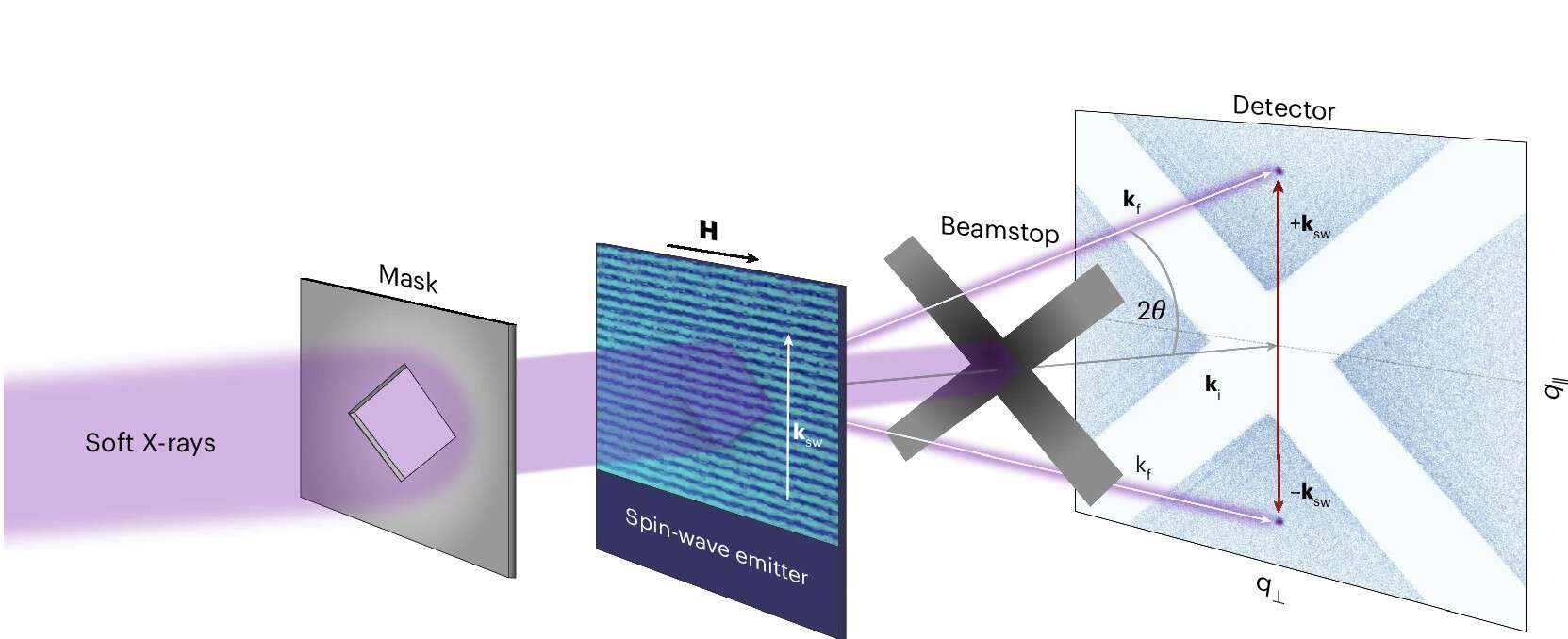
Seeing spin waves in momentum space Soft X-rays pass through a mask and scatter off spin waves travelling through a magnetic thin film (the “spin-wave emitter”). A beamstop blocks the direct beam, while the scattered X-rays reach a detector, producing two bright spots that directly reveal the spin wave’s direction and wavelength. (Courtesy: Daniel Schick/Max Born Institute/CC BY 4.0)

A new microscopy technique that can probe short-wavelength spin waves has been developed by researchers in Europe. Dubbed magnon momentum microscopy, the technique uses X-rays to observe quantized spin waves (magnons) in magnetic materials. Unlike existing techniques, the new method can characterize nonlinear interactions involving short-wavelength magnons – interactions that could be used to create new “magnonic” technologies.

The research was done by a team including Steffen Wittrock, Bastian Pfau and Daniel Schick at Germany’s Helmholtz Center Berlin for Materials and Energy and the Max Born Institute for Nonlinear Optics and Short Pulse Spectroscopy. The team comprised researchers at those German institutes and at EPFL in Switzerland.

Every ferromagnetic material is full of tiny atomic magnets, all lined up and pointing in the same direction. Nudge one of them, and the disturbance travels through the material like a ripple across a pond, passing from one atomic magnet to the next. These disturbances are called spin waves, and are quantized in units called magnons. Magnons have both particle-like and wave-like properties and physicists believe that magnons could one day carry information inside computers more efficiently than electrical signals do today. Such technologies are dubbed magnonics.

Magnon behaviour is most interesting when the magnon wavelength is shorter than about 100 nm. At these wavelengths, magnon dynamics are dominated by short-range quantum exchange interactions, rather than the longer-range forces that govern magnons with longer wavelengths.

At high magnon intensities, magnons will collide, combine, and split into new magnons before heading off in completely different directions. These nonlinear interactions will be crucial for developing magnonic technologies, but existing tools have consistently fallen short at probing this regime. Previous techniques, for example, could only probe short-wavelength magnons from one direction at a time. What magnonics researchers needed was a way to probe all directions simultaneously with extremely high sensitivity.

Now, the German–Swiss team has created an instrument that does exactly that.

Mapping in momentum space

Their technique, called magnon momentum microscopy, uses soft X-rays as a probe. When a magnon travels through a magnetic material, it leaves a regular imprint on the material’s magnetization. Soft X-rays tuned at just the right wavelength scatter off this transient imprint.

The angle of scattering directly reveals the magnon’s direction and wavelength. A detector placed behind the sample records the full scattering pattern in one shot. The result is a map in momentum space. This is a single image in which every magnon direction and wavelength appears at once, rather than one at a time. The team acquires each full map in as little as 30 s. The technique can detect magnons driven by power levels more than a thousand times weaker than what previous X-ray methods required.

Watching waves multiply

The team demonstrated the technique using a sample of yttrium iron garnet (YIG), a standard material in magnon research. They directly observed a process called four-magnon scattering, in which two high-amplitude spin waves scatter off each other and populate new waves spreading out in every direction.

On the detector, four-magnon scattering shows up as a bright elliptical ring. The ring covers all directions in 2D momentum space in a single snapshot. Its shape is a very good match for theoretical predictions of the spin-wave dispersion. Dispersion is a function that relates the propagation speed of a spin wave to the frequency of that wave.  This agreement confirms both the physics and the precision of the new technique.

At higher driving powers, new waves appear at simple fractions of the driving frequency. These fractional harmonics go beyond standard theoretical descriptions. They point to a richness in nonlinear magnon behaviour that remains to be fully understood.

With its ability to capture the full directional landscape of spin waves in one measurement, and its sensitivity reaching down to the shortest wavelengths where exchange interactions dominate, magnon momentum microscopy opens a direct route into a regime of magnon physics that was previously out of reach. The research is described in Nature Physics.

The post X-ray camera catches spin waves in the act appeared first on Physics World.

Miniature microscope lets scientists observe and control brain cells during natural movement

11 August 2026 at 20:08

A tiny microscope weighing just five grams could give neuroscientists an unprecedented view of how individual brain cells control behavior while animals move naturally. Researchers at the University of Colorado Anschutz Medical Campus and the University of Colorado Boulder have developed a head-mounted device that can both record neural activity and selectively activate individual neurons during movement. The technology could help scientists investigate how brain circuits generate behavior and how those circuits become disrupted in conditions such as Alzheimer’s disease, Parkinson’s disease and epilepsy.

The device, called Opto2P-FCM, combines two powerful techniques that have traditionally been difficult to use together in a freely moving animal. Two-photon microscopy provides detailed images of living brain tissue at cellular resolution, while optogenetics uses light-sensitive proteins to control selected neurons. By integrating both capabilities into a miniature instrument, the researchers can observe specific cells as they become active and then stimulate chosen neurons to test how they influence the surrounding network.

“These miniature microscopes have been increasingly used to study the neural basis of behavior in freely moving animals,” said Emily Gibson, PhD, associate professor of biomedical engineering at CU Anschutz and the study’s senior author. “This new microscope allows deeper penetration and higher resolution images in the brain. Importantly, it can also excite select neurons using optogenetics.”

For decades, conventional two-photon microscopes have been regarded as the gold standard for imaging neurons in living brains. These systems use infrared laser light to produce high-resolution images deep within tissue, allowing researchers to track calcium signals and other indicators of neural activity. However, conventional microscopes are generally large, stationary instruments. Animals must often remain immobilized beneath the objective, making it difficult to study brain activity during walking, exploration, social interaction or other natural behaviors.

Miniature microscopes have helped overcome that limitation by moving the imaging system onto the animal’s head. Yet reducing the size and weight of the equipment has often required compromises. Some compact systems provide lower-resolution images, while others can record neural activity but cannot stimulate precisely selected cells. The Colorado team designed Opto2P-FCM to address both problems through a dual-path optical architecture that separates imaging from stimulation.

In the imaging pathway, infrared light is directed through the brain to generate two-photon signals from fluorescent markers inside neurons. These markers can report changes in calcium concentration, which occur when neurons fire. The resulting signals are collected to create detailed images of neural activity at the level of individual cells. A separate optical pathway delivers patterned light for optogenetic stimulation, allowing researchers to activate specific neurons without disrupting the imaging beam or overwhelming the detectors.

This separation is technically important because imaging and stimulation place different demands on an optical system. High-resolution imaging requires precise focusing, efficient light collection and minimal optical distortion. Optogenetic activation, by contrast, requires intense, accurately positioned light capable of reaching the light-sensitive proteins expressed by selected neurons. Designing both functions into a device small enough to be carried by an animal required careful control of the alignment, geometry and weight of every optical component.

“This microscope is a game changer,” said Juliet Gopinath, PhD, professor of electrical, computer and energy engineering and physics at CU Boulder and a study co-author. “Being able to demonstrate a state-of-the-art instrument for both read-out and photo-stimulation of neurons is amazing.” The research team included engineers, physicists and neuroscientists who worked together to solve problems involving miniature lenses, mechanical tolerances, optical alignment and three-dimensional fabrication.

The device weighs approximately five grams, but its small size made construction unusually demanding. Mo Zohrabi, PhD, a senior research scientist at CU Boulder and co-first author, said the team initially thought such an instrument might be impossible to build because of the tight tolerances required by the optics and available 3D-printing technology. Gregory Futia, PhD, a senior research associate at CU Anschutz and co-first author, said that every component had to be designed and accounted for with extreme precision. Even slight misalignment could reduce image quality or prevent the stimulation beam from reaching the intended cells.

The researchers believe the platform could transform experiments designed to distinguish correlation from causation in the brain. Recording a neuron’s activity can show that it is associated with a behavior, but it does not prove that the cell helps produce that behavior. With optogenetic stimulation, scientists can activate a selected population and observe whether movement, decision-making or other actions change. At the same time, the microscope can record how neighboring neurons respond, providing a view of how local circuits coordinate activity.

The current Opto2P-FCM system is a prototype, and the researchers are working on future versions that could be smaller, lighter and faster. They also hope to expand the field of view so that more neurons can be monitored at once. Such improvements could make it possible to track larger networks over longer periods while animals engage in increasingly complex behaviors. By combining cellular-resolution observation with targeted manipulation in a freely moving subject, the technology may help reveal how healthy neural circuits operate and how neurological disease changes the communication between them.

Subject of Research: Miniature two-photon microscopy, optogenetics, neural circuits and brain activity during natural movement

Article Title: New Miniature Microscope Allows Scientists to Watch and Control Brain Cells During Natural Movement

News Publication Date: Aug. 11, 2026

Web References: University of Colorado Anschutz Medical Campus; University of Colorado Boulder; Optica study

References: Optica, “New Miniature Microscope Allows Scientists to Watch and Control Brain Cells During Natural Movement,” article publication date: Aug. 7, 2026

Keywords

Miniature microscope, two-photon microscopy, optogenetics, neuroscience, brain imaging, neural circuits, freely moving animals, biomedical engineering, applied physics, neurological disease, brain research

ONE Microscopy Advances High-Resolution Imaging for Scientific Discovery

11 August 2026 at 13:54

Expansion microscopy has spent the past decade changing the rules of super-resolution imaging. Instead of relying solely on increasingly sophisticated optics, the technique physically enlarges biological specimens so that molecules separated by nanometers become easier to distinguish with ordinary fluorescence microscopes. Now, researchers have introduced a method designed to push that concept toward one of the most difficult goals in microscopy: seeing the shapes of individual proteins directly.

The approach, described by Ahmed H. Shaib, Mahmoud M. Alawieh and Stefan O. Rizzoli in Nature Protocols, combines one-step nanoscale expansion microscopy with fluctuation-based super-resolution analysis. The resulting workflow, called ONE microscopy, is intended to make molecular-scale imaging more accessible to laboratories that do not have access to cryo-electron microscopes or specialized high-end optical systems.

Traditional light microscopy is limited by diffraction, a physical effect that causes light from two nearby objects to blur together when they are too close. Even modern super-resolution methods can require complex instruments, intense labeling strategies or extensive computational processing. Expansion microscopy takes a different route. Researchers anchor biological molecules within a swellable polymer network and then expand the material, increasing the physical distance between fluorescent labels.

The expansion step does not automatically reveal every molecular detail. A protein’s structure can still be represented by only a small number of fluorescent signals, and conventional images may contain noise, background fluorescence and motion-related fluctuations. ONE microscopy addresses these limitations by analyzing changes in fluorescence intensity over time. These fluctuations contain information about the presence, position and behavior of labeled molecules that may not be obvious in a single frame.

In practical terms, the method links chemical preparation, physical enlargement and computational analysis into a single workflow. Samples are embedded in a polymer gel, labeled to identify the proteins of interest and then expanded. After expansion, researchers acquire image sequences using conventional fluorescence equipment. Specialized software analyzes the temporal variation in the recorded signals, extracting spatial information beyond what a standard diffraction-limited image would provide.

This combination is significant because it shifts the focus from simply locating a protein to examining its overall shape. For many biological questions, knowing that a protein is present is not enough. Its size, orientation and structural organization can determine how it interacts with membranes, vesicles, organelles or neighboring proteins. Directly observing those features could help researchers investigate molecular machines in their native cellular environments rather than relying exclusively on purified samples or averaged structural models.

The protocol is designed to work across a broad range of biological materials, including purified proteins, cultured cells and tissues. That flexibility could make the technique useful for researchers studying protein organization at multiple scales. A purified protein might provide a controlled test of shape reconstruction, while cells and tissues could reveal how the same protein is arranged amid the crowded and complex environment of living biology.

A major part of the reported advance is the accompanying software package. Fluctuation-based imaging can be powerful, but its usefulness depends on reliable data processing, and computational analysis has often been a barrier for non-specialist users. The authors present the software as stable and user-friendly, with the goal of making the analysis more efficient, reproducible and practical for laboratories using standard fluorescence microscopes.

ONE microscopy does not replace cryo-electron microscopy, which remains capable of resolving structures at atomic or near-atomic scales under appropriate conditions. Nor does it eliminate the challenges associated with labeling, gel chemistry, image quality and sample preparation. Expansion can introduce distortions, and successful imaging depends on preserving the relationship between the fluorescent labels and the underlying structures. Nevertheless, the method offers a complementary strategy: rather than averaging thousands or millions of molecules, it aims to examine individual protein shapes through expanded, fluorescence-labeled specimens.

The researchers describe the workflow as a practical framework for protein imaging on conventional equipment. By combining physical separation of fluorophores with information extracted from fluorescence fluctuations, the method brings nanoscale structural analysis closer to routine biological imaging. Its broader impact may come not from replacing existing forms of super-resolution, but from making a previously specialized capability more reproducible and attainable for laboratories investigating how individual proteins operate inside cells and tissues.

Subject of Research: One-step nanoscale expansion microscopy for visualizing individual protein shapes using conventional fluorescence microscopes.

Article Title: ONE microscopy.

Article References: Shaib, A.H., Alawieh, M.M. & Rizzoli, S.O. ONE microscopy. Nature Protocols (2026). https://doi.org/10.1038/s41596-026-01399-x

Image Credits: AI Generated

DOI: https://doi.org/10.1038/s41596-026-01399-x

Keywords: expansion microscopy, ExM, nanoscale imaging, super-resolution microscopy, fluctuation-based analysis, protein structure, fluorescence microscopy, single-protein imaging, cryo-electron microscopy, biological imaging

Frost spreads across surfaces via suspended ‘ice bridges’

9 July 2026 at 10:17

Frost can spread not only along surfaces but also via suspended “ice bridges” that form above them. The discovery of this previously unknown frost-propagation pathway could lead to new ways of making surfaces that resist frost growth, thereby improving the performance of devices that operate in cold, humid environments.

Frost accumulation is a major problem in refrigerators, aeroplanes and heat pumps, to name just a few. On the microscale, it primarily spreads from one individual freezing water droplet to the next via two-dimensional bridges, or causeways, that temporarily form on the surface of an object. Although the wettability of the surface is known to affect the spreading process, the mechanism behind this effect was poorly understood.

Ice bridges can grow in two distinct spatial modes

To learn more, a team led by physicist Nenad Miljkovic at the University of Illinois Urbana-Champaign, US imaged the channel-forming process using high-speed high-resolution optical microscopy combined with a profilometry technique called focal plane shift imaging (FPSI). They found that frost can spread in two distinct ways. On hydrophilic surfaces, causeways form along the substrate, in line with current theoretical models. On superhydrophobic surfaces, however, the situation is quite different. Here, frost spreads via ice bridges that are suspended above the surface in three-dimensional space.

This suspended or “out-of-plane” growth mode represents a fundamentally different pathway for frost propagation, says team member Siyan Yang, the first author of a Nature Physics  paper about the work. Previous studies likely overlooked this mechanism, Yang adds, because of limitations in experimental observations.

Superhydrophobic coatings nearly double the frost propagation time

The researchers also studied the growth rate of the different bridge types. They found that suspended bridges grew slower than bridges on the surface due to the reduced thermal coupling between the bridges and the cold substrate.  This reduced coupling correspondingly reduces the vapour pressure difference between ice and water droplets (which depends on surface droplet geometry, itself controlled by wettability) and drives down ice growth. Indeed, the team found that the speed at which frost spreads fell more than 80% in this mode.

To test the practical relevance of their findings, the researchers applied superhydrophobic coatings to metre-sized structures such as the finned-tube aluminium heat exchangers commonly found in air conditioners, refrigerators and automotive systems. Condensation frosting on such systems poses a major efficiency challenge because frost has an inherently low thermal conductivity. When it accumulates on heat exchangers, it therefore severely impedes heat being exchanged with the surrounding air.

On uncoated commercial heat exchangers that are inherently hydrophilic, the team found that frost rapidly forms and spreads across the fins. “In the superhydrophobic counterparts, however, the onset of frost formation is delayed, and it propagates much more slowly,” Yang says. In the two kinds of commercial systems they tested, she adds, applying superhydrophobic coatings nearly doubled the frost propagation time.

The results suggest that designers of anti-frost surfaces could benefit from trying this new strategy, Yang says. “Rather than focusing solely on delaying initial ice nucleation, surfaces could be engineered to control the geometry of ice-bridge growth and interrupt frost spreading, thereby improving the performance and energy efficiency of a host of equipment operating in cold and humid environments,” she tells Physics World.

The team is now investigating how surface chemistry and surface structures influence suspended ice-bridge formation and frost propagation. “We are also exploring ways to translate the fundamental mechanism into scalable anti-frost coatings and heat-exchanger technologies,” Yang reveals. “Ultimately, our goal is to establish predictive design rules that connect microscale ice-bridge dynamics with real-world frost management performance.”

The post Frost spreads across surfaces via suspended ‘ice bridges’ appeared first on Physics World.

Laser-based rotation of floating samples enables high-resolution 3D microscopy

18 June 2026 at 10:00

A new contact-free way of rotating delicate samples, such as biological cells, undergoing three-dimensional (3D) optical microscopy increases the image resolution and could reveal more details about microscale structures and processes.

Current optical microscopes provide extremely good image resolution in a single plane, but since depth information is often lacking, samples are routinely rotated and imaged at multiple different angles to create a 3D picture. Because rotating via mechanical microtools such as tiny grippers, needles or pipettes can damage samples, contact-free approaches such as optical tweezers, magnetic manipulation or optothermal forces are often used instead. However, these methods rely on the sample being birefringent, magnetic, light-absorbing or specially shaped.

By contrast, the laser-based technique developed by a team led by Moritz Kreysing and Fan Nan from the Institute of Biological and Chemical Systems at the Karlsruher Institut für Technologie (KIT) in Germany can rotate and spin floating microscopic objects no matter what their shape or material properties.

Their method involves focusing a 1455 nm infrared laser beam on a small region of a highly viscous liquid – initially at 25°C – in which the sample will be suspended. This gently heats that region of the liquid by a few degrees. But rather than focusing on one spot, an acousto-optic deflector steers the beam rapidly along a carefully designed two-dimensional (2D) pattern.

“Because the viscosity of the liquid changes with temperature, this moving heat pattern generates tiny fluid flows, known as thermoviscous flows. By choosing the scan geometry and timing correctly, these flows become three-dimensional and helical, like microscopic corkscrew-shaped currents,” explain Kreysing and Nan. Once a small object is floated freely in these 3D helical flows, it can be rotated, spun, transported or stabilized solely via software-defined scan patterns, with different symmetries controlling the movement of the surrounding liquid.

The KIT technique allows stacks of high-resolution images focused at different depths to be acquired along multiple different angles by re-orienting the sample, a feature that “can help overcome the anisotropic resolution of conventional 3D microscopy”, explain the researchers. Using a high-viscosity liquid to suspend the samples not only enables kinematic stop-and-go actuation, it also suppresses Brownian motion (the jiggling around of tiny particles in fluids), which otherwise decreases image resolution and makes the study of cellular transport processes tricky.

Since previous methods of laser-driving fluid flows could only create motion in one plane, the researchers found it “particularly exciting” when they first observed their laser scanning in a 2D plane generating 3D flow fields. “Another key moment was the observation of opto-hydrodynamic focusing. The particles did not just rotate; their spiral motion converged toward a stable height and position. This was significant because it meant that the optofluid could stabilize the sample, rather than requiring mechanical confinement or complex feedback control.”

As detailed in their recent Light: Science & Applications paper, the KIT team demonstrated rotation and spinning of various samples including stained biological cells, nano-printed micro-tiles and perfectly spherical beads. “We see immediate potential in advanced optical microscopy, especially multi-view imaging of suspended cells, cell clusters, organelles, microstructures or soft materials,” Kreysing and Nan tell Physics World.

Since the basic hardware components of their system will be familiar to many optics and microscopy labs, they are hoping for early adoption by specialized research groups. But to optimize the technique for biological applications, they will now investigate the use of different liquids and operating conditions “that better preserve the viability of live mammalian cells while still providing sufficient stability for precise rotation and imaging”.

The researchers suggest that future areas of application could include microfluidics, microrobotics, colloidal assembly, materials science and 3D microfabrication. As such, they also want to develop more advanced scan patterns that can generate curved, programmable or multi-axis helical flows. “This could allow more complex 3D manipulation, such as controlled orientation of irregular objects, coordinated actuation of multiple particles, or integration with microfabrication workflows,” they explain.

The post Laser-based rotation of floating samples enables high-resolution 3D microscopy appeared first on Physics World.

Scientists Have Identified a Key Driver of Age-Related Cognitive Decline

Dementia Diagnostics MRI Brain ScanA newly identified protein pathway may link blood-brain barrier deterioration to age-related cognitive decline. The aging brain depends on a microscopic border that works every second to keep danger out and support the nerve cells inside. When that barrier begins to fail, memory, mood, and thinking may suffer. Andrew A. Pieper, MD, PhD, and colleagues [...]

Stanford’s Revolutionary New Microscope Reveals Living Cells in Stunning Detail

31 May 2026 at 21:31
Interferometric Microscopy Laser ArrayStanford researchers have developed a microscope that can show how nanostructures interact inside living cells at the highest resolution achieved so far. The view into living cells just got better. Stanford researchers have merged two microscopy methods to build a unique instrument that can capture cell structures interacting in real time at an unprecedented resolution [...]
❌