❌

Normal view

Open-source software tracks wrist bone motion in 3D and 4D CT scans

9 September 2026 at 18:20

In a development that could reshape how scientists study the mechanics of human movement, a team of biomedical engineers and computer scientists has unveiled an open-source software tool capable of tracking the tiny, intricate bones of the wrist as they glide and rotate during motion. The new tool, called Hierarchical 3D Registration, or 3DH, is a module within the SlicerAutoscoperM extension of the widely used 3DSlicer computing environment. Described in a study published in BioMedical Engineering OnLine, the software was validated against previously established methods using both sequential three-dimensional computed tomography, or 3DCT, scans of living participants performing thumb tasks and dynamic four-dimensional CT, or 4DCT, images of cadaveric wrists moving through flexion and extension. The results demonstrate that the open-source approach can match the accuracy of independent, previously published methodologies while offering researchers everywhere a free and reproducible path to quantifying skeletal kinematics.

The wrist has long been one of the most challenging joints for biomechanists to study. It contains eight small carpal bones, including the scaphoid and lunate, along with the radius of the forearm and the metacarpal bones of the hand, all articulating in a tightly packed arrangement that allows the remarkable range of motion humans rely on every day. Understanding how these bones move relative to one another, a field of study known as arthrokinematics, is essential for unraveling the causes of wrist instability, osteoarthritis, ligament injuries and the long-term outcomes of surgical repairs. Yet measuring those movements in living, moving joints has historically required either invasive techniques, labor-intensive manual analysis or proprietary software that few laboratories can access or verify.

Quantitative tracking of skeletal structures in vivo, with both efficiency and high temporal resolution, matters to a wide range of questions in biomechanical research, and the authors of the new study argue that the field has been held back by a shortage of accessible, semi-automated registration tools. Image registration, the computational process of aligning one three-dimensional image volume to another, lies at the heart of motion tracking from CT data. When a bone moves between two scans, its position and orientation change, and a registration algorithm must find the transformation that best overlays the bone’s image from one time point onto the next. Done well, this yields precise measurements of rotation and translation. Done poorly or inconsistently, it introduces errors that can swamp the small movements of carpal bones, which may shift by only a few millimeters and a few degrees during normal function.

The new 3DH module addresses this problem with a hierarchical strategy for registering bones across sequences of image volumes. The researchers presented and evaluated the software using two distinct classes of data drawn from previous studies. The first consisted of sequential 3DCT datasets of human participants performing various thumb tasks, in which the radius, the first metacarpal and the trapezium were the target bones. The second consisted of dynamic 4DCT datasets capturing simulated flexion-extension motion in cadaveric wrists, providing time-resolved, three-dimensional image volumes of the scaphoid and lunate in motion. By testing the tool on both static sequential scans and true dynamic imaging, the team was able to demonstrate that 3DH is not limited to a single imaging paradigm but can serve as a unified tracking solution across modalities.

To determine whether the new open-source tool could be trusted, the investigators compared the arthrokinematics computed with 3DH against values that had been previously calculated using independent approaches for the same 3DCT and 4DCT datasets. The statistical framework for this comparison was the Bland-Altman analysis, a standard method for assessing agreement between two measurement techniques. Rather than simply correlating the two sets of results, Bland-Altman analysis quantifies the mean bias, the systematic difference between methods, and the 95 percent limits of agreement, the range within which most individual differences between the methods are expected to fall. This approach is particularly well suited to method-comparison studies because it reveals whether a new technique introduces systematic error and how widely individual measurements might deviate from those of the established gold standard.

The performance figures reported in the study are striking. For the 3DCT data, in which the radius, first metacarpal and trapezium were tracked, the mean bias for the helical angle, a single-axis description of three-dimensional rotation, was just minus 0.06 degrees, with 95 percent limits of agreement ranging between minus 1.87 degrees and 1.74 degrees. Translation measurements fared nearly as well: the mean bias was 0.12 millimeters, with limits of agreement spanning from minus 2.23 millimeters to 2.00 millimeters. In practical terms, the open-source tool reproduced the rotational and translational measurements of the earlier methods to within roughly two degrees and two millimeters at the extremes, differences small enough to preserve the biological signal in most wrist kinematics experiments.

The dynamic 4DCT results told a similar story for the carpal bones. Tracking the scaphoid and lunate through simulated flexion-extension, 3DH produced mean biases of the Euler angle rotations, which decompose three-dimensional rotation into three orthogonal components, of less than 0.77 degrees about any direction. The widest 95 percent limits of agreement ranged between minus 7.11 degrees and 8.65 degrees. For translations, mean biases were less than 0.5 millimeters in any direction, with the widest limits of agreement ranging between minus 2.36 millimeters and 1.43 millimeters. The wider limits of agreement for rotations in the dynamic data likely reflect the added challenges of time-resolved imaging, including motion blur and the smaller, more complexly shaped carpal bones, but the near-zero mean biases indicate that the tool does not systematically drift away from established values.

Crucially, all target bones were successfully tracked in both the 3DCT and 4DCT datasets, meaning the hierarchical registration algorithm converged on correct solutions in every case tested. This robustness matters because registration algorithms can occasionally fail, locking onto incorrect alignments or diverging entirely, and such failures typically demand time-consuming manual intervention. A semi-automated tool that reliably succeeds across diverse bones, motion tasks and imaging protocols removes a significant bottleneck. Manual bone tracking in CT volume sequences can take hours per bone per trial, and the labor multiplies rapidly in studies involving multiple participants, multiple bones and dozens of motion cycles. By automating this process within a validated framework, 3DH promises to accelerate the pace at which wrist biomechanics data can be collected and analyzed.

The choice to build the module inside 3DSlicer, and specifically within the SlicerAutoscoperM extension, is a deliberate strategic decision with implications well beyond the wrist. 3DSlicer is a free, open-source platform for medical image computing and visualization with a large international user base spanning research, clinical and educational communities. By embedding 3DH in this ecosystem, the developers have made the tool immediately available to any laboratory already working with 3DSlicer, complete with the platform’s established infrastructure for loading DICOM data, segmenting anatomical structures and visualizing results. The authors note that the ability to track bones from both 3DCT and 4DCT data sources, with agreement to prior methodologies, demonstrates that the 3DH approach is a capable tool that could promote collaboration and consistency of results through open-source software.

Consistency is a quietly urgent issue in biomechanics. When different laboratories use different proprietary tools with undisclosed algorithms, it becomes difficult to distinguish true biological differences between study populations from mere methodological discrepancies. Open-source registration changes that calculus: the code can be inspected, verified, extended and reused, and results from different institutions become directly comparable because they are produced by identical algorithms. This transparency also lowers the barrier to entry for research groups in settings that cannot afford commercial licenses, potentially broadening the global community capable of conducting rigorous skeletal kinematics research.

The study itself emerged from a collaboration spanning several institutions. The team included Joseph J. Crisco, Amy M. Morton and John D. Holtgrewe of the Department of Orthopedics at The Warren Alpert Medical School of Brown University and Rhode Island Hospital; Cesar Lopez, Andrew Thoreson and Kristin D. Zhao of the Assistive and Restorative Technology Laboratory and the Department of Physical Medicine and Rehabilitation at the Mayo Clinic in Rochester, Minnesota; and Jean-Christophe Fillion-Robin and Beatriz Paniagua of Kitware Inc. in Clifton Park, New York. The involvement of Kitware, a company known for its leadership in open-source scientific computing, underscores the blend of academic rigor and software engineering expertise required to produce a research tool robust enough for widespread distribution. Funding was provided by the National Institute of Arthritis and Musculoskeletal and Skin Diseases of the National Institutes of Health under award numbers AR059185, AR078924 and AR071338.

The implications of this work extend naturally to the future of musculoskeletal imaging. As newer CT technologies, including photon-counting detector systems, deliver ever-finer spatial resolution and as dynamic 4DCT becomes more widely available, the volume of image data available for kinematic analysis will grow dramatically. Tools like 3DH, which are fast, semi-automated and validated across modalities, will be essential for converting that imaging bounty into quantitative biomechanical knowledge. For patients with wrist disorders, the downstream promise is equally tangible: better measurements of how carpal bones actually move in living joints can inform more accurate diagnoses, improved surgical planning, more realistic computational models of joint loading and ultimately better-designed treatments for conditions ranging from scapholunate instability to thumb-base arthritis. With the tool now openly available in the 3DSlicer environment, researchers around the world can begin putting it to work.

Subject of Research: Open-source kinematic tracking of the small bones of the wrist in sequential 3DCT and dynamic 4DCT images using Hierarchical 3D Registration

Article Title: Kinematic tracking of the small bones of the wrist in sequential 3DCT and dynamic 4DCT volume images using open-source Hierarchical 3D Registration, a module within SlicerAutoscoperM

Article References: Crisco, J. J., Morton, A. M., Lopez, C., Thoreson, A., Zhao, K. D., Holtgrewe, J. D., Fillion-Robin, J.-C., & Paniagua, B. (2026). Kinematic tracking of the small bones of the wrist in sequential 3DCT and dynamic 4DCT volume images using open-source Hierarchical 3D Registration, a module within SlicerAutoscoperM. BioMedical Engineering OnLine. https://doi.org/10.1186/s12938-026-01601-x

Image Credits: AI Generated

DOI: 10.1186/s12938-026-01601-x

Keywords: Computed tomography, 3DCT, 4DCT, Bone tracking, 3DH, 3DSlicer, Image registration, Wrist kinematics, Arthrokinematics, Open-source software, SlicerAutoscoperM, Bland-Altman analysis

Cardiac Breakthrough Could Offer Lifesaving Treatment to More Patients

8 September 2026 at 22:34
Healthy Human Heart AnatomyA major trial found that implantable cardioverter-defibrillators may benefit some patients younger than 70 with mild-to-moderate heart dysfunction and cardiac scarring. A more than decade-long randomized trial suggests that implantable cardioverter-defibrillators may benefit some patients who have heart scarring and only mild-to-moderate loss of heart function, a group that currently falls outside standard treatment guidelines. [...]

ONE Microscopy Advances High-Resolution Imaging for Scientific Discovery

11 August 2026 at 13:54

Expansion microscopy has spent the past decade changing the rules of super-resolution imaging. Instead of relying solely on increasingly sophisticated optics, the technique physically enlarges biological specimens so that molecules separated by nanometers become easier to distinguish with ordinary fluorescence microscopes. Now, researchers have introduced a method designed to push that concept toward one of the most difficult goals in microscopy: seeing the shapes of individual proteins directly.

The approach, described by Ahmed H. Shaib, Mahmoud M. Alawieh and Stefan O. Rizzoli in Nature Protocols, combines one-step nanoscale expansion microscopy with fluctuation-based super-resolution analysis. The resulting workflow, called ONE microscopy, is intended to make molecular-scale imaging more accessible to laboratories that do not have access to cryo-electron microscopes or specialized high-end optical systems.

Traditional light microscopy is limited by diffraction, a physical effect that causes light from two nearby objects to blur together when they are too close. Even modern super-resolution methods can require complex instruments, intense labeling strategies or extensive computational processing. Expansion microscopy takes a different route. Researchers anchor biological molecules within a swellable polymer network and then expand the material, increasing the physical distance between fluorescent labels.

The expansion step does not automatically reveal every molecular detail. A protein’s structure can still be represented by only a small number of fluorescent signals, and conventional images may contain noise, background fluorescence and motion-related fluctuations. ONE microscopy addresses these limitations by analyzing changes in fluorescence intensity over time. These fluctuations contain information about the presence, position and behavior of labeled molecules that may not be obvious in a single frame.

In practical terms, the method links chemical preparation, physical enlargement and computational analysis into a single workflow. Samples are embedded in a polymer gel, labeled to identify the proteins of interest and then expanded. After expansion, researchers acquire image sequences using conventional fluorescence equipment. Specialized software analyzes the temporal variation in the recorded signals, extracting spatial information beyond what a standard diffraction-limited image would provide.

This combination is significant because it shifts the focus from simply locating a protein to examining its overall shape. For many biological questions, knowing that a protein is present is not enough. Its size, orientation and structural organization can determine how it interacts with membranes, vesicles, organelles or neighboring proteins. Directly observing those features could help researchers investigate molecular machines in their native cellular environments rather than relying exclusively on purified samples or averaged structural models.

The protocol is designed to work across a broad range of biological materials, including purified proteins, cultured cells and tissues. That flexibility could make the technique useful for researchers studying protein organization at multiple scales. A purified protein might provide a controlled test of shape reconstruction, while cells and tissues could reveal how the same protein is arranged amid the crowded and complex environment of living biology.

A major part of the reported advance is the accompanying software package. Fluctuation-based imaging can be powerful, but its usefulness depends on reliable data processing, and computational analysis has often been a barrier for non-specialist users. The authors present the software as stable and user-friendly, with the goal of making the analysis more efficient, reproducible and practical for laboratories using standard fluorescence microscopes.

ONE microscopy does not replace cryo-electron microscopy, which remains capable of resolving structures at atomic or near-atomic scales under appropriate conditions. Nor does it eliminate the challenges associated with labeling, gel chemistry, image quality and sample preparation. Expansion can introduce distortions, and successful imaging depends on preserving the relationship between the fluorescent labels and the underlying structures. Nevertheless, the method offers a complementary strategy: rather than averaging thousands or millions of molecules, it aims to examine individual protein shapes through expanded, fluorescence-labeled specimens.

The researchers describe the workflow as a practical framework for protein imaging on conventional equipment. By combining physical separation of fluorophores with information extracted from fluorescence fluctuations, the method brings nanoscale structural analysis closer to routine biological imaging. Its broader impact may come not from replacing existing forms of super-resolution, but from making a previously specialized capability more reproducible and attainable for laboratories investigating how individual proteins operate inside cells and tissues.

Subject of Research: One-step nanoscale expansion microscopy for visualizing individual protein shapes using conventional fluorescence microscopes.

Article Title: ONE microscopy.

Article References: Shaib, A.H., Alawieh, M.M. & Rizzoli, S.O. ONE microscopy. Nature Protocols (2026). https://doi.org/10.1038/s41596-026-01399-x

Image Credits: AI Generated

DOI: https://doi.org/10.1038/s41596-026-01399-x

Keywords: expansion microscopy, ExM, nanoscale imaging, super-resolution microscopy, fluctuation-based analysis, protein structure, fluorescence microscopy, single-protein imaging, cryo-electron microscopy, biological imaging

Biomedical Imaging Foundation Models: Separating Hype from Reality

11 August 2026 at 13:48

Foundation models are promising to become the “universal translators” of biomedical data—but a new perspective argues that medicine should resist treating them as all-knowing digital doctors. In a review published in Nature Biomedical Engineering, researchers describe how large, adaptable artificial intelligence systems are reshaping biomedical imaging while warning that impressive benchmark scores can conceal serious weaknesses in clinical environments. Their central message is direct: foundation models are most likely to improve healthcare by augmenting specialists, not replacing them.

Foundation models are designed to learn broad representations from enormous and diverse datasets before being adapted to specific tasks. In biomedical imaging, those data may include magnetic resonance imaging, computed tomography, X-rays, ultrasound, digital pathology slides and ophthalmic images. The same underlying model could theoretically identify tumors, segment organs, estimate disease risk, retrieve similar cases and generate clinical reports. The vision becomes even broader when imaging is combined with pathology, electronic health records and genomic information, creating a composite system intended to analyze a patient across multiple biological scales.

That ambition reflects a major change from traditional medical AI. Earlier systems were generally built for one narrowly defined task, such as detecting pneumonia on chest radiographs or outlining a brain lesion on MRI scans. Foundation models instead attempt to create a reusable backbone that can be transferred across diseases, hospitals and imaging technologies. Technically, they learn statistical patterns in high-dimensional data, often through self-supervised training in which the system predicts missing, transformed or associated information. Afterward, developers fine-tune or prompt the model for particular clinical applications.

But biomedical imaging is not a single, uniform world. Images differ according to scanner manufacturer, acquisition protocol, patient population, disease prevalence and local clinical practice. A model trained primarily on data from large academic hospitals may perform very differently in community clinics, rural settings or countries with limited equipment. This problem, known as domain shift, can occur even when images appear visually similar. Small changes in image quality, contrast, hardware or patient demographics may alter the statistical distribution learned by the model and undermine its predictions.

The authors introduce a framework called real-world evaluation and assessment of foundation models, or REAL-FM, to examine whether these systems are ready for practical use. Rather than focusing only on accuracy scores, REAL-FM considers several dimensions at once, including the quality and representativeness of training data, technical readiness, clinical value, integration into existing workflows and responsible artificial intelligence. The framework is intended to help clinicians interpret claims about new models and to push developers toward evaluations that resemble actual medical practice.

One of the sharpest distinctions in the perspective is between pattern recognition and causal reasoning. Foundation models can become extraordinarily skilled at recognizing visual associations—for example, linking a particular texture or anatomical feature with a diagnosis present in their training data. Yet association is not the same as understanding why a disease occurs, how it will progress or whether a treatment will benefit an individual patient. A model may identify a correlation that is valid in one hospital but reflects a hidden confounder, such as a scanner type, reporting convention or patient-selection pattern, rather than a biological signal.

This limitation becomes especially dangerous when models are moved beyond simplified benchmarks. Many benchmark datasets provide carefully curated images, clear labels and narrowly defined tasks. Clinical care is messier: scans can be incomplete, diagnoses can be uncertain, records may contain contradictory information and several conditions may coexist. The perspective highlights a lack of verified generalization across such conditions, along with a shortage of prospective, outcome-based validation. In a prospective study, an AI system would be evaluated while care is actually being delivered, with researchers measuring whether it improves diagnostic accuracy, treatment decisions, patient outcomes or workflow efficiency.

Data scarcity is another obstacle. The largest foundation models require vast quantities of data, but medical information is difficult to collect, standardize and share. Patient records are fragmented across institutions, imaging data may lack reliable annotations and rare diseases are inherently underrepresented. Privacy regulations and governance requirements further complicate the creation of centralized datasets. As a result, a model may appear broadly capable while remaining poorly tested in the populations and clinical circumstances where errors could have the greatest consequences.

The researchers argue that human oversight therefore remains indispensable. In practical terms, foundation models may be most useful as clinical assistants that prioritize images for review, highlight suspicious regions, summarize longitudinal records or offer a second opinion that specialists can interrogate. Safe deployment will require transparent reporting of uncertainty, monitoring for performance drift, mechanisms for correcting errors and clear accountability when recommendations influence care. The future envisioned by the authors is not a single monolithic medical oracle, but a coordinated ecosystem of specialized AI systems, each evaluated for a defined clinical role and connected to expert-led workflows.

The perspective arrives as the biomedical AI field races toward increasingly general-purpose systems. Its warning is not that foundation models lack value, but that technical scale alone cannot establish clinical reliability. Before these models can be trusted with consequential decisions, they must demonstrate robustness across domains, usefulness in real workflows, safety under unexpected conditions and measurable benefits for patients. The proposed REAL-FM framework offers a way to separate viral demonstrations from durable medical progress—by asking not only what a model can recognize, but where it works, why it works and whether it makes care better.

Subject of Research: Foundation models and their real-world evaluation in biomedical imaging and clinical medicine.

Article Title: Foundation models in biomedical imaging: turning hype into reality

Article References: Muneer, A., Zhang, K., Hamdi, I. et al. Foundation models in biomedical imaging: turning hype into reality. Nature Biomedical Engineering 10, 1557–1575 (2026). https://doi.org/10.1038/s41551-026-01762-z

Image Credits: AI Generated

DOI: 10.1038/s41551-026-01762-z

Keywords: foundation models, biomedical imaging, medical artificial intelligence, clinical AI, multimodal AI, domain shift, causal reasoning, clinical validation, responsible AI, REAL-FM

Schizophrenia’s Brain Damage May Start in One Key Region

10 August 2026 at 20:25
Woman Mental Health Depression Anxiety SchizophreniaSchizophrenia may leave a distinctive biological footprint across the brain’s communication network. Using specialized PET imaging, researchers have mapped where synaptic connections are most reduced in people with schizophrenia and identified a possible region from which the damage begins to spread. The study, which involved a Rutgers professor, was published in Molecular Psychiatry. Synapses are [...]

Webb Found a Hidden Planet in Beta Pictoris By Reading Its Atmosphere

9 August 2026 at 04:00

Astronomers have found a third giant planet around Beta Pictoris, one of the best-known young planetary systems near Earth. The discovery is striking because the planet was not first picked out as a bright dot in a telescope image. NASA’s James Webb Space Telescope found it in the spectrum of light coming from the system: […]

The post Webb Found a Hidden Planet in Beta Pictoris By Reading Its Atmosphere appeared first on Curiosmos.

Europe's free satellite service just made it easier to track wildfires

7 August 2026 at 22:48

The wildfires afflicting communities across the world can now be tracked more easily by anyone using Europe’s free and open satellite imagery service—all thanks to a wildfire visualization script originating from nearly a decade ago.

The Copernicus Browser provides free access to the latest satellite imagery taken by the European Space Agency’s Sentinel satellites for the European Union’s Earth observation program. The three satellites of the Sentinel-2 mission have proven especially useful for wildfire monitoring by providing multi-spectral imaging at resolutions as granular as 10 meters in visible light and near-infrared bands—and the Copernicus Browser previously offered 10 default visualization layers using different combinations of spectral bands.

Now Copernicus Browser has also added a straightforward “wildfires” visualization layer for Sentinel-2 imagery that went live on August 4. Anyone clicking on this layer while looking at a wildfire location can see active fires in white or yellow, burning vegetation in red, and burned landscapes in dark brown or black.

Read full article

Comments

© European Union | Copernicus Sentinel-2

Portable breast ultrasound enables imaging between cancer screening exams

3 August 2026 at 10:00
<em>In vivo</em> ultrasound images
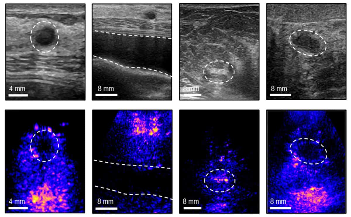
In vivo ultrasound images From left to right: a cyst, breast implant, dense fibrous tissue and solid mass. The top row shows conventional 2D ultrasound images while the bottom row shows the corresponding images using the 3D PURE system. (Courtesy: the researchers)

Women at high risk of breast cancer are more susceptible to developing cancers in the gap between routine screening exams. These so-called “interval cancers” tend to be fast-growing and account for 20% to 30% of all breast cancer cases.

To identify aggressive cancers that develop between scheduled screenings, researchers at Massachusetts Institute of Technology (MIT) have developed a point-of-care ultrasound system that generates high-resolution 3D images of breast tissue. The 3D portable ultrasound system for real-time examination (3D PURE) supplements mammography with its ability to detect anomalies reliably in dense breast tissue (in which mammography is less sensitive).

Guided by an easy-to-use visual interface, 3D PURE enables a nurse, a physician such as a primary care provider or gynaecologist, or even the patient herself to frequently perform longitudinal breast imaging.

Second-generation design

Principal investigator Canan Dagdeviren explains that this second-generation prototype overcomes limitations of a prior system designed by the MIT team, through advances in transducer design, acoustic materials and adaptive beamforming. These developments have enabled real-time wide-angle 3D imaging in a portable form factor, with improved image resolution to accurately identify cysts, solid masses, fibroadenomas and microcalcifications.

The ultrasound system comprises a newly designed 128-element box-shaped 2D transducer array incorporating a corner-gap offset geometry, which suppresses peak crosstalk by 3.73 dB at the corner-most element. This design prevents amplifier saturation, and supports operational transmit voltages of up to 24 V. The array is integrated into a compact multilayer electronics stack comprising preamplifiers, transmit electronics and a custom chirp data acquisition system.

There’s also a conductive backing layer (a toluene-diluted polyurethane matrix with a high percentage of tungsten and zirconia filler) that forms a continuous shielding layer around the elements, improving acoustic attenuation and providing electromagnetic shielding. The compliant mechanical nature of the material enhances acoustic damping, reducing ultrasound reverberations more effectively and improving signal fidelity.

“The addition of a backing layer to the ultrasound transducer is a key advantage to the system,” explains co-lead author Md Osman Goni Nayeem. “It provides both better directionality of ultrasound waves and better bandwidth, which improves the resolution and quality of the resulting images.”

The researchers also incorporated layered aberration-correction reconstruction (LACR), an adaptive 3D beamformer, into the 3D PURE design. LACR compensates for the heterogeneous speed-of-sound in the breast, which comprises glandular and fibrous tissues plus a superficial layer of fat. They believe that this represents the first wide-angle 3D ultrasound imaging system to implement aberration-correction beamforming.

“What we are trying to do is predict the speed-of-sound properties of the tissue being imaged, and then use that to reconstruct the image more accurately. We see up to a 10% improvement for the resolution just by applying this [beamforming] technique,” says co-lead author Shrihari Viswanath in a press statement.

A 3D ultrasound probe positioned on a tissue phantom
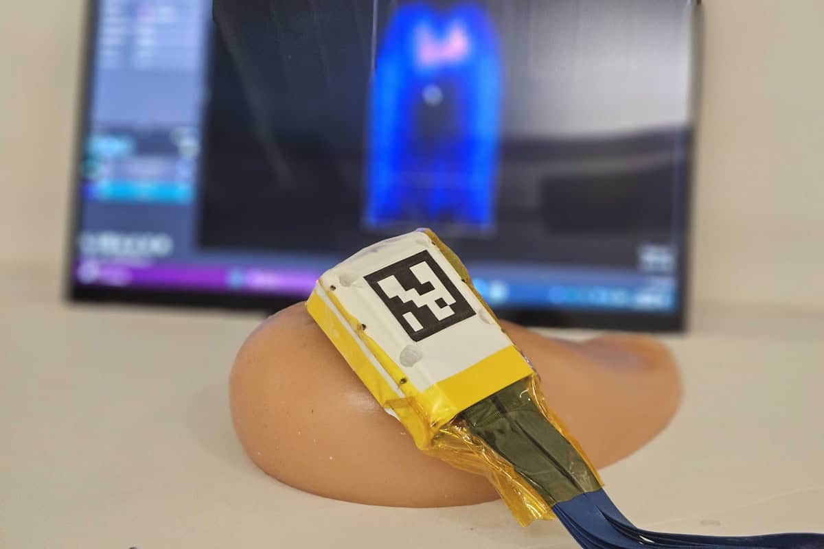
Experimental validation The 3D ultrasound probe is positioned on a tissue-mimicking breast phantom for visualization of embedded microtargets. (Courtesy: the researchers)

The system is designed for ease of use. A visual user interface called “Mirror my First UltraSound” (MyFUS) guides users to reliably reposition the ultrasound probe at the same anatomical location during repeated imaging and long-term monitoring. Meanwhile, a wide field-of-view minimizes the number of scans needed to cover the entire breast.

The team undertook an in vitro study comparing the performance of the 3D PURE system with that of a conventional 2D handheld ultrasound system (HHUS). Ten study participants imaged a breast-shaped tissue phantom embedded with sub-millimetre targets. Nine of the 10 participants showed improved microtarget detection efficiency with 3D PURE relative to the conventional system – identifying more of the targets using 3D PURE (79.7%, compared with 60.7% for the 2D HHUS), with a higher detection efficiency (14.02% of available targets per minute, compared with 9.58%).

The researchers also worked with radiologists to assess a variety of breast anomalies in vivo. The radiologists verified that the 3D PURE system could accurately visualize calcifications, cysts, implants, fibrous tissues and solid masses within a large volumetric field-of-view. The researchers independently validated the system’s accuracy when imaging dense fibrous breast tissue and rib structures.

Seven volunteers with no prior experience of using an ultrasound system tested the MyFUS visual interface. After initial instruction by an ultrasound technician, they were able to repetitively position the probe at a specific location on their breasts, achieving a 94.10% mean overlap value on frontal projections, and 87.42% on side projections. This test reconfirmed that novice users could achieve reproducible longitudinal monitoring results with the aid of the visual interface.

The researchers now hope to create an interface for use with mobile phones or tablets. This type of system could make breast ultrasound more accessible to patients in economically constrained countries lacking adequate breast cancer scanning and regions with shortages of trained ultrasound technicians.

“The high operator dependence of conventional ultrasound is a barrier to its use in decentralized or home-monitoring settings,” the researchers write. “The MyFUS vision interface transforms longitudinal monitoring from an expert-driven procedure to a self-guided, reproducible process that can be used by novice users. This reproducibility is vital for longitudinal monitoring in limited clinical settings, to regularly track the growth of an anomaly or the response of a tumour to therapies without visiting clinics.”

The 3D PURE system is described in Nature Communications.

The post Portable breast ultrasound enables imaging between cancer screening exams appeared first on Physics World.

AI sniffs out tuberculosis in breath’s chemical signature

31 July 2026 at 12:00

Every breath we exhale carries a complex chemical narrative written in thousands of volatile organic compounds (VOCs). These tiny molecules, produced by human metabolic processes and invading pathogens, hold immense diagnostic potential. Yet, translating this molecular cloud into a definitive medical diagnosis has long faced an analytical bottleneck. Now, an international team of physicists from Botswana and South Africa has harnessed machine learning to decode these complex signatures, offering a rapid, non-invasive pathway to detect tuberculosis (TB) directly from human breath.

Breaking the analytical bottleneck

Traditional gas chromatography-mass spectrometry (GC-MS) excels at separating complex molecular mixtures based on volatility and mass-to-charge ratios. However, a single breath sample generates thousands of overlapping spectral peaks. Manually sifting through this dense landscape is a painstaking, time-consuming process.

To find a faster, more systematic approach to molecule identification, researchers in the Nano –Breath taking group led by George Chimowa from Botswana International University of Science and Technology (BIUST) collected breath samples from three distinct cohorts: patients with active TB, individuals with multidrug-resistant TB (MDR-TB), and healthy control volunteers.

Standard analytical pipelines often rely on aggressive data pre-processing to smooth and simplify the GC-MS spectra. However, this filtering risks erasing subtle, low-concentration molecular features that might hold critical diagnostic clues. Bypassing this limitation, the BIUST group fed a high-dimensional dataset of 1867 spectral features (from 87 breath samples) directly into four competing supervised machine-learning algorithms: decision trees, random forest, k-nearest neighbours, and support vector machines (SVM). The researchers published their findings in Discover Artificial Intelligence.

Geometric optimization avoids the pitfalls of high-dimensional data

The team found that the linear SVM classifier led the algorithmic comparison, distinguishing among the three patient cohorts with 93% accuracy.

This performance stems from how the algorithm handles high-dimensional data spaces. While distance metric models often suffer from overfitting errors, where experimental noise is misidentified as a meaningful diagnostic signal, SVM relies on geometric boundary optimization to separate classes.

Much like clearing a wide highway down the centre of two opposing crowds, it identifies a hyperplane that maximizes the margin between groups. Instead of tracking every individual in the crowd, the algorithm relies solely on a sparse subset of border points known as support vectors. By focusing strictly on these boundary “guards” and ignoring the background movement, this approach completely isolates the classifier from the noise inherent to the surrounding high-dimensional feature space.

The Nano – Breath taking research group

Beyond raw classification accuracy, the model yielded a crucial insight for future clinical applications. It pinpointed a specific window within the gas chromatogram, a retention time between 10 and 30 min, where the most diagnostic, high-variance VOCs elute. This 20-min window corresponds to medium- and large-molecule VOCs, including specific chemical biomarkers associated with TB, such as tridecane, decane and O-cymene.

By demonstrating that these vital diagnostic features are concentrated within this single slice of data, the physicists have shown that future diagnostic hardware can be specialized, eliminating the need to analyse the entire spectrum. Narrowing the focus to this specific elution window can drastically shrink data-processing times and maximize instrument throughput in clinical settings.

AI and the future of medical diagnostics

TB remains one of the world’s deadliest infectious diseases. Traditional diagnostics rely heavily on slow sputum cultures and backlogged laboratory queues, delaying critical treatment. A rapid, non-invasive breath test powered by an optimized physical classifier offers an ideal alternative for resource-limited settings where laboratory infrastructure may be scarce.

While the researchers emphasize that larger validation trials are required, the work signals a profound shift in medical diagnostics. It mirrors advanced data-modelling techniques used in engineering, such as those predicting structural degradation in self-healing aerospace composites.

By fusing analytical physics with artificial intelligence, this research points to a future where immediate, automated bedside disease detection could be rapid, accessible and driven entirely by the mathematical profiling of a patient’s breath.

The post AI sniffs out tuberculosis in breath’s chemical signature appeared first on Physics World.

5,000-Year-Old Blue Pigment Reveals Evidence of Ancient Egyptian Nanotechnology—Now It’s Finding New Life in Biomedical Imaging

28 July 2026 at 14:05

Thousands of years ago, a striking blue pigment began appearing in the art that adorned the walls of ancient Egyptian tombs, and eventually on the crown of the famous bust of Nefertiti, wife of the 18th dynasty pharaoh Akhenaten and half-sister to one of Ancient Egypt’s most famous kings: Tutankhamun.

However, the ancient artists who first used this remarkable pigment, known today as “Egyptian blue,” had likely never guessed that thousands of years into the future, this unique substance would have applications in modern biomedical research.

That is precisely what one team of researchers discovered several years ago, in a study that revealed how this striking pigment is more than just a modern medical marvel with origins in the ancient world—it also provides evidence of ancient nanotechnology in early Egypt.

The Enigma of Egyptian Blue

“Egyptian blue” is the common term for a pigment first developed in ancient Egypt around 2500 B.C., made from calcium copper silicate. Originally, it was created to provide an artificial alternative to a more costly form of blue pigment from the era, which was produced using lapis lazuli.

However, ancient Egyptian scientists discovered that by heating limestone, sand, and copper, they could produce a far less expensive form of blue pigment, and soon its use spread throughout the ancient Mediterranean world, making its way into the craftsmanship in parts of the ancient world that ranged from Mesopotamia to Greece and Rome.

An Ancient Technology Finds Modern Biomedical Applications

Although Egyptian blue’s obvious practical uses in art were derived from its unique and stunning appearance, time would eventually reveal that it had other uses. Specifically, in a 2020 study published in Nature Communications, a team of researchers reported on their discovery that Egyptian blue in its powdered form can be separated into nanoscale sheets roughly 100,000 times thinner than a human hair.

This is significant because these tiny particles of the substance exhibit significant fluorescence under near-infrared light, which can be used by researchers to track them within biological samples.

Chemist Sebastian Kruss of Georg-August University Göttingen, the lead author of the 2020 study, remarked at the time that he was certain such capabilities “will be of growing interest for material scientists as well as biomedical research” in the years ahead.

Ancient Nanotech’s Survival Through the Centuries

One reason the pigment is so useful is also the likely reason behind its discovery, and that is Egyptian blue’s durability. For many millennia, samples of this unique material have managed to survive, and the material’s nanoscale form remains stable even over such long periods.

In the past, earlier studies had demonstrated that the material fluoresces in near-infrared wavelengths. Additionally, it was known to scientists that the pigment could be broken down into flakes after being stirred in hot water for periods of several days.

Building on this knowledge, the research detailed in the 2020 paper sought to go even further by examining whether those particles could function as fluorescent markers in living systems.

Near-Infrared Applications

One thing that scientists find useful about near-infrared imaging is that it provides an effective way to examine biological material by leveraging wavelengths that fall just beyond the ranges of the visible spectrum.

Although some molecules can already be used as fluorescent markers, Egyptian blue nanoparticles generate significantly brighter signals than several common alternatives. Additionally, unlike other fluorescent compounds, the pigment can resist photobleaching, which is important because this allows its glow to persist even during prolonged observation.

A pair of experiments was used by the team to demonstrate the material’s potential. The first involved using Egyptian blue particles injected within a fruit fly embryo, which were tracked as it developed. In another, the particles were added to plant leaves, allowing comparisons with the performance of a conventional near-infrared imaging molecule.

In this experiment, the team found that the Egyptian blue produced a bright signal that remained visible even against the leaves’ natural fluorescence—the comparison molecule, by contrast, was heavily obscured.

According to Kruss, the tiny Egyptian Blue particles prove to be “very useful glowing labels in biomedical research,” calling the pigment a “very powerful fluorophore.”

An Ancient Discovery Breaking New Ground in the 21st Century

According to Kruss and his colleagues, such particles could eventually help scientists observe processes including embryonic development and cell division, while potentially supporting applications such as image-guided surgery and the labeling of cancerous tissue.

Altogether, this enigmatic form of ancient nanotechnology is still playing a role in helping develop the cutting-edge biomedical technologies of tomorrow, even more than 5,000 years after its initial discovery.

Kruss and his team’s original paper from 2020, “Exfoliated near infrared fluorescent silicate nanosheets for (bio)photonics,” was published in Nature.

Micah Hanks is the Editor-in-Chief and Co-Founder of The Debrief. A longtime reporter on science, defense, and technology with a focus on space and astronomy, he can be reached at micah@thedebrief.org. Follow him on X @MicahHanks, and at micahhanks.com.

Shaping the field of medical imaging: a fireside chat with Simon R Cherry and Xinchen Deng

23 July 2026 at 14:47


Join us for an in‑depth conversation with Simon Cherry, former editor‑in‑chief of Physics in Medicine & Biology (PMB), as he shares his expert perspective on the evolving landscape of medical physics research, molecular imaging, and non-invasive imaging technologies. Drawing on decades of leadership in biomedical imaging and academic publishing, Simon will reflect on major advances shaping the field, define the characteristics of high‑impact imaging research, and highlight how PMB continues to support rigorous, influential scientific work through its editorial standards and peer‑review practices.

This session will additionally spotlight the research of Xinchen (Alison) Deng, a finalist of the PMB 2024 Early Career Award. Attendees will gain insights into her contributions to the advanced medical imaging community, such as photon-counting CT for clinical applications and how her work supports broader advances in medical physics and X-ray/CT imaging research.

Simon will also discuss what makes early‑career research stand out and share strategies for researchers aiming to increase the visibility, reach, and long‑term impact of their publications within competitive scientific journals.

Carol Clark, Simon Cherry, Xinchen Deng
Left to right: Carol Clark, Simon Cherry, Xinchen (Alison) Deng

Moderator

Carol Clark, Publisher, Physics in Medicine & Biology

Speakers

Simon R Cherry, PhD received his BSc (Hons) in physics with astronomy from University College London in 1986 and a PhD in medical physics from the Institute of Cancer Research, University of London in 1989. Simon is currently Distinguished Research Professor in the Departments of Biomedical Engineering and Radiology at UC Davis and Professor of Total-Body Imaging at King’s College London. Simon’s research interests centre around biomedical imaging. His major contributions have been in developing systems for positron emission tomography (PET), including designing the microPET scanner for small-animal imaging, the earliest examples of simultaneous PET and MRI studies and co-leading the EXPLORER consortium that developed the world’s first total-body PET scanner. Simon is a Fellow of the Royal Society and Member of the U.S. National Academy of Engineering and National Academy of Inventors. He has received several international awards including the Society of Nuclear Medicine and Molecular Imaging Cassen Prize in 2022. He served as editor-in-chief of the journal Physics in Medicine & Biology from 2011–2020. Simon is the author of more than 350 peer-reviewed journal articles, review articles and book chapters in the field of biomedical imaging. He is also lead author of the widely-used textbook Physics in Nuclear Medicine.

Xinchen (Alison) Deng, PhD, is a diagnostic imaging physics resident at The Ohio State University. Her research focuses on advancing photon-counting CT for clinical applications, including multi-contrast imaging, early disease detection, detector optimization, and AI-based improvements in image quality. Before beginning the residency, Xinchen (Alison) was a postdoctoral fellow in medical physics at the University of Victoria, where she worked with Magdalena Bazalova-Carter and collaborated with Redlen Technologies on the optimization of photon-counting detectors and the clinical applications of spectral CT. She received her PhD in medical physics from the University of British Columbia, where she applied Raman spectroscopy, machine learning, and explainable AI to study radiation treatment response in cancer cells and tissues. Her broader research background spans photon-counting CT, spectral X-ray imaging, Raman spectroscopy, radiotherapy response assessment, and AI-driven image and data analysis. Her photon-counting CT research has been featured in the Physics in Medicine & Biology Early Career Researcher Focus Collection. She is a finalist for the PMB Early Career Researcher Award.

About this journal

Physics in Medicine & Biology. The international journal of biomedical physics and engineering, published by IOP Publishing on behalf of the Institute of Physics and Engineering in Medicine (IPEM)

Editor-in-chief: Katia Parodi, Ludwig-Maximilians University, Munich, Germany


The post Shaping the field of medical imaging: a fireside chat with Simon R Cherry and Xinchen Deng appeared first on Physics World.

Privacy risks from medical AI are greater than previously thought

21 July 2026 at 15:30
Schematic of a membership inference attack
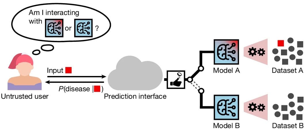
Membership inference attack An untrusted user aims to infer whether a specific target record was part of the training dataset, by observing the predictions of a model. The attack is successful if they can reliably distinguish between models A and B, which are identical except for the inclusion or exclusion of the target record in the respective training dataset. (Courtesy: CC BY 4.0/Nature 10.1038/s41586-026-10688-0)

A new study has revealed that sensitive health information about individual patients can be extracted from medical artificial intelligence (AI) models more effectively than previous research had indicated.

Diagnostic AI models are used to determine whether some given health data belong to a specific class – for example if an X-ray image shows evidence of pneumonia – and offer huge promise for improving patient outcomes. Earlier studies showed that membership inference attacks (MIAs), which determine whether a particular person’s data was used to train an AI model, yielded results no better than random chance. But these previous studies only evaluated the average risk across all patients in a training dataset.

By contrast, in this latest work doctoral student Moritz Knolle and his supervisor Daniel Rückert at the Technical University of Munich (TUM), together with collaborators at Imperial College London and Georg Kaissis from the Hasso Plattner Institute (HPI), examined the privacy risk for an individual patient. “Privacy that only works on average has little meaning. So I asked myself: would I contribute my data to train a medical AI model?” says Knolle.

Daniel Rückert from TUM
Investigating the risks A team led by Daniel Rückert (pictured) at TUM and Georg Kaissis from HPI found that medical AI is more vulnerable to certain attacks than previously thought. (Courtesy: Juli Eberle/TUM)

As detailed in their recent Nature paper, the researchers attacked AI models based on seven different types of real-life medical datasets, including one comprising 21,799 electrocardiograms and another of 377,110 chest radiographs.

“If a model sees a specific data point during training, it usually becomes more confident at predicting it. So in these MIA attacks I compare the predictive probability number for this data point outputted by the medical AI model with the predictive probability number obtained from my model which has not seen that data point,” explains Knolle, adding that the attack is essentially a statistical test to distinguish between the two models.

Their results revealed that the privacy risk for an individual was far greater than the average risk. The team also found that privacy risk is not equally distributed among subgroups.

“We found that patients from minority groups that are under-represented in the training dataset tend to be over-represented among the 99 percentile of extreme [privacy] risk,” Knolle tells Physics World, warning that as medical AI models become larger and more sophisticated the privacy risk is amplified because their deep analysis creates the small changes in predictive probability that MIAs are good at finding.

While an MIA that reveals someone is a UK citizen poses little security risk, discovering that an individual has received a specific type of cancer treatment reveals personal information that could, for instance, adversely affect their medical insurance premiums. “The more specific the training cohort is, the larger the privacy risk,” states Knolle, crediting his medical doctor parents with inspiring his desire to improve healthcare AI.

Countermeasures include differential privacy (DP), in which a small amount of Gaussian random white noise is added in alongside the data during training, thereby reducing the effectiveness of MIAs while maintaining the model’s diagnostic accuracy. “This gives a mathematically provable upper bound for how much privacy risk any individual in this dataset could face, by limiting the information content that flows to the AI model,” explains Knolle.

He notes that, unfortunately, such DP tools have not been widely adopted by hospitals or clinics. This, he fears, could undermine patients’ confidence in sharing their data, and thereby not only reduce AI’s ability to help combat rare diseases, but also compound AI discrimination that occurs when there is too little data from minority groups included in training datasets.

“So we are hoping to inspire the medical AI community to do risk assessments before deploying AI models, and use verifiable strategies such as DP or limiting access to the AI model, so that as an individual you have a statistical guarantee that no harm would come to you by being part of a dataset,” concludes Knolle, who now plans to investigate the privacy risks posed by generative AI models for healthcare.

The post Privacy risks from medical AI are greater than previously thought appeared first on Physics World.

First medical X-ray images taken in space achieve diagnostic quality

15 July 2026 at 09:35

A collaboration of researchers in North America has reported their findings from the first diagnostic X-ray imaging performed in space. X-ray images recorded during an orbital spaceflight exhibited similar overall quality to those taken preflight on Earth, demonstrating the feasibility of adding radiography as a diagnostic tool for crew health.

With humans venturing further into space and spending more time in this harsh environment, the risk of adverse medical events increases. Accurate and timely diagnosis and treatment is a must-have. Currently, the only reliable medical imaging modality available to astronauts in spaceflight is ultrasonography – which is operator dependent, requires substantial training and relies on limited acoustic windows.

X-ray imaging, on the other hand, benefits from low operator dependence, rapid acquisition and typically higher spatial resolution than ultrasound. Digital radiography could also offer superior diagnostic capabilities for many medical conditions of concern during spaceflight, including dental disease, musculoskeletal trauma, inhalational injury, collapsed lung and arthritis.

Traditional X-ray scanning systems are bulky, produce a lot of radiation and are sensitive to motion – making them less than ideal for sending into space. But a new generation of portable X-ray scanners shows potential to survive the spaceflight and be operated by crew members with minimal training.

“It’s been a dream for aerospace medicine to have more than one imaging modality for diagnosing illnesses and injuries in space,” says lead researcher Sheyna Gifford, an assistant professor of aerospace medicine at the Mayo Clinic in Rochester, MN, in a press statement.

Scanning in space

In a study led by co-author David Lerner, the researchers first demonstrated the feasibility of X-ray imaging in simulated microgravity in 2022. During a parabolic flight, they used a commercial off-the-shelf digital radiography system to successfully capture radiographs of a human subject and a phantom during lunar gravity (1/6g), Martian gravity (1/3g) and microgravity.

In this latest project, reported in in Radiology, Gifford and her team investigated the performance of a similar portable digital radiography system aboard the Dragon spacecraft, during the Fram2 spaceflight mission, a 3.5-day polar orbital flight launched on 31 March 2025 on a SpaceX Falcon 9 rocket. The imaging system comprised a commercial off-the-shelf, FDA-cleared, portable, digital X-ray generator (Impact Wireless; MinXray) along with an FDA-cleared flat-panel detector.

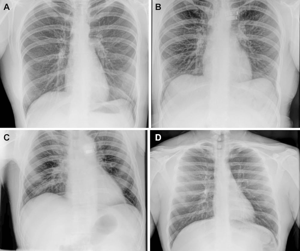
Chest radiographs Representative X-ray images acquired preflight by a crewmember (A), in-flight on day 3 after launch by a crewmember (B, C) and postflight by a non-crew operator using the same imaging protocol (D). (Courtesy: RSNA)

Prior to the mission, three crew members received 4 hr of training on the portable X-ray system and acquired preflight X-ray images of the hand, forearm, abdomen, pelvis, chest and a quality control phantom. They then recorded the same anatomic and phantom images whilst in space, and also imaged a smartwatch to investigate the potential for non-destructive testing.

During anatomic imaging, the devices were handheld by the crew members, while the phantom and smartwatch were secured to the detector for imaging. The crew reported that they found it easy to use the X-ray system and follow the imaging protocols.

Back on Earth

To assess the quality of the seven X-ray images acquired by the crew, three independent radiologists evaluated and compared the preflight and in-flight anatomic radiographs, finding no differences in overall image quality, contrast resolution or spatial resolution. For central-body radiographs (chest, abdomen and pelvis), image positioning was worse in-flight than preflight. Despite this, all X-rays achieved good or near-excellent diagnostic quality (rated on a  5-point Likert scale).

Phantom radiographs recorded in space exhibited good spatial and contrast resolution, imaging low-contrast targets down to the smallest 2-mm target and easily visualizing high-contrast meshes ranging from 20 to 60 lines per inch, with 80 lines per inch remaining visible. The smartwatch radiograph clearly showed the watch’s internal components at the submillimetre scale.

The research team found that the X-ray generator had sustained minor re-entry damage, but its internal components and X-ray output were not affected. Similarly, the detector passed visual inspection and quality control tests. The researchers also recorded a set of postflight X-rays (on non-crewmembers) following the same in-flight protocols. These showed no qualitative differences from the preflight or in-flight radiographs.

“By acquiring the first human and equipment X-rays in space, our study demonstrates the feasibility of in-orbit radiography and expanded diagnostic capabilities for crew health and hardware evaluation,” says Gifford. “Acquiring diagnostically useful X-rays in space is something that anyone can do. Three very talented nonmedical people with four hours of training in one of the harshest environments did it right and did it well.”

Gifford points out that a spaceflight-ready radiography system could also prove indispensable for mission-critical non-medical tasks. “For sustained human presence in space, X-rays are critical not just for crew members but also for other mission components like electronics and spacesuits. The only way to look inside these objects without taking them apart is to X-ray them,” she explains.

The post First medical X-ray images taken in space achieve diagnostic quality appeared first on Physics World.

Mouldable glass screen could make mammograms clearer and much more comfortable

29 June 2026 at 14:00

A new glass screen containing nanoclusters of copper iodide could create higher-resolution X-ray images using a much lower dose of radiation. The scintillating screen, which also works underwater, can be moulded into curved shapes, overcoming key limitations of the conventional rigid, flat-panel detectors used for many medical imaging applications. These include mammography, say the researchers at King Abdullah University of Science and Technology (KAUST), Saudi Arabia, and the University of Hamburg, Germany, who developed it.

X-ray scintillators capture X-rays passing through an object and convert them into flashes of visible light to form a digital image, explains physicist Mehmet Bayindir of KAUST and the University of Hamburg. The more efficient the scintillator is at this conversion, the clearer the final image and the lower dose of radiation required to generate it.

He and his colleagues’ detailed analyses of a scintillator containing nanoclusters of copper, iodine and an organic ligand revealed a fascinating phenomenon: its photoluminescence and radioluminescence pathways appear to have distinct physical origins. This implies that the scintillating light yield – that is, how much visible light a material emits when struck by high-energy X-rays – can be separated from its standard photoluminescence quantum yield (how efficiently the material converts absorbed ultraviolet or visible light into a different wavelength of light).

Until now, Bayindir explains, it was assumed that a material must have high photoluminescence efficiency to perform well when excited by X-rays. “Breaking this assumption directly challenges conventional wisdom and opens up a promising new design space for making high-performance scintillators – provided that materials highly responsive to radiation can be engineered of course.”

Enter amorphous quantum systems

In recent years, researchers have been looking into cubane-type [Cu4I4L4, L: organic ligand] nanocluster glasses (NCGs) for use in this context. These belong to a family of hybrid transparent luminescent amorphous materials that show much promise for X-ray scintillation applications.

In their latest work, detailed in ACS Energy Letters, Bayindir and colleagues studied several stable alkyl-phosphine ligand-based NCGs. In these amorphous zero-dimensional quantum systems, ionizing radiation is mostly absorbed by the heavy iodine and copper elements at the cluster cores. The researchers used these NCGs to make large-area, free-standing scintillating screens – both planar and conformal – with ultrasmooth surfaces, using a fabrication methodology that they developed in their lab.

The screens are robust and can image samples – both electronic and biological – with sub-3 µm spatial resolution. They also work underwater, something that is difficult for conventional screens. The researchers demonstrated this by capturing a very clear scan of a fish’s tail in water that was indistinguishable from an image taken in air.

Such a glass scintillator represents a significant milestone, says team leader Osman Bakr. “Indeed, the level of precision we obtained allows us to capture highly detailed, high-contrast X-ray images of complex internal microstructures, such as the delicate anatomy of a microscopic insect and an electronic memory card, to cite but two examples,” he says.

X-ray imaging systems that curve to fit a person’s body

“Combined with the fact that this new nanocluster glass becomes almost rubbery when heated to 42 °C, we can mould it into any three-dimensional shape at low temperatures,” study lead author Bashir Hasanov adds. “This could allow for X-ray imaging systems that curve to fit a person’s body and overcomes the limitations of conventional rigid, flat-panel detectors.”

One possible application is in mammography, he notes. Today, a major drawback of current mammography machines is that breast tissue must be firmly compressed between flat panels so that it can be imaged. This is very uncomfortable – and even painful – and deters many women from going for a scan.

“One in 20 women around the world will be diagnosed with breast cancer in their lifetime, with annual cases projected to reach 3.2 million and related deaths climbing to 1.1 million by 2050,” says Bayindir. “Many of these fatalities stem from late-stage diagnosis.

Nearly one-third of women miss their initial mammogram appointments, something that significantly increases their risk of succumbing to the disease. “While the reasons for this vary, the medical literature frequently cites the physical discomfort of tissue compression,” says Bakr. “What is more, because current guidelines recommend starting routine mammography screenings at age 40, cumulative lifetime exposure to X-ray radiation remains a clinical concern for many. Our long-term objective is to completely redefine this clinical experience.”

Indeed, the researchers say they would now like to bring this quantum glass out of the lab and into real-world applications. Bakr tells Physics World that his team’s immediate next step is to scale up the geometric dimensions of its mouldable screens while maintaining perfect thickness and clarity. “This would move us closer to full-scale clinical prototyping for 3D conformal medical imaging.”

“We are also designing a novel detector architecture capable of high-resolution imaging on curved surfaces using an array of specialized optical sensors,” he reveals.

The post Mouldable glass screen could make mammograms clearer and much more comfortable appeared first on Physics World.

Affordable AI-powered endomicroscope lines up for early cancer detection

26 June 2026 at 09:00

Epithelial cancers account for 80% to 90% of all cancer cases in the world, comprising a wide variety of malignancies that begin in the epithelial tissues that form the skin and line the body’s internal passageways and organs. Early diagnosis is critical to improving patient outcomes, but is limited by a heavy reliance on invasive biopsies.

Simultaneous assessment of changes in both the cell nuclei and the microvasculature could potentially improve the accuracy of early cancer and precancer detection, and help guide decisions regarding biopsy and pathology testing. In vivo microscopy (IVM) has shown promise for aiding early diagnosis of epithelial cancers, but has challenges relating to image quality, spatial resolution and field-of-view (FOV).

PrecisionView, an artificial intelligence (AI)-powered, dual-modality endomicroscope in development at Rice University’s Rice360 Institute for Global Health Technologies, addresses these IVM issues. The compact, handheld, easy-to use system enables real-time, wide-area visualization of both subcellular structures and underlying blood vessels, offering higher-quality imaging, a fivefold increase in FOV, and an eightfold increase in spatial resolution over endomicroscopic systems in current clinical use.

The proof-of-concept version of PrecisionView, described in Proceedings of the National Academy of Sciences, is intended for superficial epithelial imaging where scattering-induced distortions remain moderate. And it is specifically designed to be low cost: providing an affordable diagnostic tool for clinics worldwide.

PrecisionView uses simple, off-the-shelf miniaturized optics integrated with a deep learning-based optimization framework. Its AI-supported phase mask design and reconstruction algorithm incorporate dual-modality fluorescence and reflectance imaging to achieve a 5.2 x 3.9 FOV and a 500 µm depth-of-field (DOF) while maintaining cellular-level resolution (4 µm). Images are acquired at approximately 15 frames/s, in 2 s intervals, with image tiles stitched together to form a large-scale, co-registered map of cell nuclei and microvasculature.

Performance assessment

To validate the system, principal investigators Rebecca Richards-Kortum and Ashok Veeraraghavan, and collaborators at the University of Texas MD Anderson Cancer Center, analysed images of ex vivo porcine tongue, human breast and fresh cervical tissue specimens with precancerous lesions.

High-resolution maps of cervical specimens containing squamous intraepithelial lesions (precancer) clearly delineated key anatomic structures, including columnar and squamous epithelium, and the junction between the two epithelial cell types. PrecisionView clearly distinguished abnormal regions from surrounding benign tissue – suggesting that the system may be able to support physicians’ decisions regarding whether or not to conduct a biopsy.

The researchers also performed real-time, in vivo imaging of the oral cavities of healthy volunteers. They report that the system’s large DOF enabled acquisition of high-resolution images of cell nuclei and microvasculature across a large tissue area without requiring refocusing. When the mucosal surface was scanned, the large FOV of each frame, combined with the high video acquisition rate, provided substantial frame-to-frame overlap. This allowed seamless stitching of the dual-modality images to create large-scale, co-registered maps of the oral mucosa, which showed distinct cellular and vascular features.

Meeting a global need

The Rice 360 Institute focuses on designing, implementing and scaling highly effective, low-cost medical technologies for underserved and low-resource communities. The idea for PrecisionView grew out of recognizing a global limitation in access to high-quality diagnostic information.

“In many care settings, especially where conventional pathology resources are limited, clinicians may not have timely access to the cellular- and tissue-level information needed to guide diagnosis and treatment decisions,” says first author Huayu Hou, who worked with co-first author Jimin Wu, to develop the PrecisionView instrument.

Hou explains that although existing clinical endomicroscopy systems can provide valuable microscopic information, their broader use is often limited by cost, complexity, a limited imaging volume and the ability to capture only a narrow range of diagnostic features. Such constraints reflect fundamental design trade-offs in compact clinical optical imaging systems and create significant barriers to practical point-of-care use.

“PrecisionView was motivated by the opportunity to use computational imaging to overcome limitations in existing clinical endomicroscope designs that could not be fully addressed with traditional optical approaches alone, with the goal of bringing high-quality diagnostic information closer to the point-of-care,” he says.

Hou advises that it took about a year to develop the first working prototype, and that several projects are now underway to further enhance both performance and usability for point-of-care applications. “We are working to improve image quality and resolution, so that the system can provide finer cellular details from living systems to real time,” he adds. “We are working to further refine the hardware and workflow to make the device easier to operate in clinical settings.”

The team is developing AI-based classification algorithms that can integrate both cellular and vascular information and provide real-time diagnostic guidance. Its long term goal is to create a system that not only visualizes tissue at the cellular level, but also helps clinicians interpret the information efficiently at the point-of-care. “This capability could be especially valuable in low-resource settings, where access to pathology expertise may be limited, and tools that support real-time decision-making could significantly improve clinical usability and impact,” notes Wu.

Clinical validation studies to evaluate PrecisionView’s diagnostic performance across different clinical settings and cancer types are also planned. For instance, a study focusing on head-and-neck cancer detection has already started in collaboration with MD Anderson Cancer Center. Others are being developed for cervical cancer imaging in Mozambique, to evaluate the system’s performance in low-resource settings.

The post Affordable AI-powered endomicroscope lines up for early cancer detection appeared first on Physics World.

X-ray velocimetry study earns Ronan Smith the PMB Early Career Researcher Award

4 June 2026 at 09:00

Ronan Smith, a postdoctoral research fellow at Adelaide University, has been chosen as the winner of this year’s Physics in Medicine & Biology (PMB) Early Career Researcher Award. The award is presented to the author of the “best paper” in PMB’s 2025 Early Career Researcher Focus Collection, as selected by the journal’s editorial board.

Smith’s research involves implementing X-ray velocimetry (XV), a novel imaging method that uses X-rays to track lung motion during breathing and create 3D maps of local ventilation. In his award-winning paper, Visualising ventilation changes following endobronchial valve placement with x-ray velocimetry functional lung imaging, Smith investigates the potential of XV imaging to detect changes in lung function after insertion of an endobronchial valve (EBV).

EBVs are one-way valves that are placed into the lung to help treat emphysema – a condition that damages air sacs in the lungs, causing air to get trapped inside and making breathing difficult. The EBV, which in some cases can be used in place of surgery, prevents airflow into damaged lung areas so that the rest of the lung can function more effectively.

Successful valve placement causes the targeted area of lung to collapse, which can be imaged using CT. Smith proposed that use of XV functional lung imaging to non-invasively measure regional and local changes in airflow could more accurately assess the clinical impact of EBV placement.

“The lungs are a dynamic organ, their job is to be constantly moving,” he explains. “Because X-ray velocimetry looks at lung motion, it lets us see exactly where the air is or isn’t flowing, so you can instantly see that airflow has changed within the lungs. CT only measures structural changes, which may not necessarily be correlated to changes in lung function.”

In vivo demonstration

To investigate this potential advantage, Smith and colleagues carried out a pilot study on healthy sheep, which have a similar lung size to humans. They performed XV imaging on two anaesthetized and ventilated animals, before and after placing EBVs in their lungs.

The XV scanning process involves recording fluoroscopic videos of individual breaths at various angles around the lung, with anatomic positioning provided by an accompanying breath-hold CT scan. To analyse the data, the researchers used XV LVAS software from 4DMedical, the MedTech company that developed and commercialized the XV technology.

XV and CT imaging
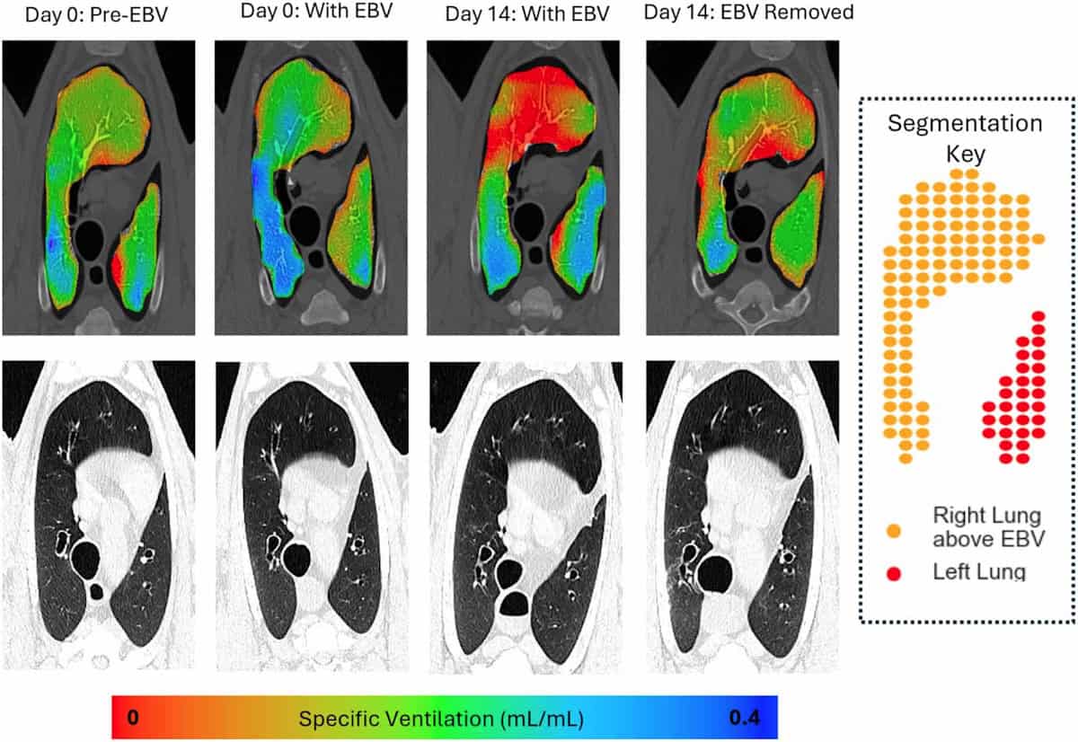
Visualizing ventilation Slices from XV and CT imaging taken from an area of lung above the EBV. XV shows clear changes in ventilation immediately after valve insertion. (Courtesy: Phys. Med. Biol. 10.1088/1361-6560/ade196)

The software correlates motion in the XV videos with CT data to measure the lung’s expansion and contraction during a breath cycle. It creates a 3D map of specific ventilation (the change in voxel volume during a breath, divided by its starting volume) in small voxels throughout the lungs. This map can then be used to calculate mean specific ventilation and ventilation heterogeneity across a given lung region.

As soon as the EBVs were inserted into the animals’ lungs, XV imaging could visualize and quantify a reduction in airflow to areas downstream of the valves. This effect was seen both in regions where collapse was visible in CT scans, as well as those where collapse could not be detected by CT. Ventilation changes were also clearly observed in the remainder of the lungs.

“The main finding of our study was that X-ray velocimetry imaging can detect airflow changes from endobronchial valve placement in the lungs,” says Smith. “Our research could be really important for people [with emphysema], as tools to help with better placement and verification will lead to improved treatment options.”

Future prospects

Since the publication of this paper, Smith has focused on further applications of pre-clinical and clinical XV imaging. “I’ve been working as part of a great interdisciplinary team looking at how lung function changes in a range of diseases, to both understand the diseases, and as an outcome measure when we test treatments,” he says.

This work includes the world’s first paediatric clinical trial of XV imaging, which is examining the feasibility of using the technology in children with cystic fibrosis. The researchers have imaged around 30 children to date and aim to publish their findings later this year. They are currently planning future studies to see how XV imaging could enhance clinical decision making and improve outcomes for these children, as well as looking at other childhood diseases where it could be of relevance.

“As an early-career researcher, I’m also focussing on developing my own research, looking at another novel X-ray imaging method called dark-field X-ray imaging,” Smith adds.

The perfect award

Smith tells Physics World that he was excited to receive the PMB Early Career Researcher Award, acknowledging the efforts of everyone involved in this “hugely collaborative project”, including clinicians, scientists, 4DMedical and the staff in the preclinical imaging facility where the study was performed.

Ronan Smith and colleagues
Team effort Ronan Smith (back row, centre) and colleagues in the lab. (Courtesy: Women’s and Children’s Health Network South Australia)

“As a physicist working in medicine/biology, it feels like the perfect award to get,” he says. “It’s great to see interest in the work we are doing, and fantastic evidence we can use to convince the funding bodies it’s worth continuing this work.”

  • The PMB Early Career Researcher Focus Collection 2025 publishes papers from early-career researchers (defined for this collection as a postgraduate student or someone who completed their PhD in 2019 or later) in biomedical physics, with the aim of highlighting research excellence from emerging leaders. The Early Career Researcher Award was introduced to further recognise an outstanding contribution from one of the early-career authors, selected according to the quality of scientific content, number of citations and downloads, and peer review ratings.

The post X-ray velocimetry study earns Ronan Smith the PMB Early Career Researcher Award appeared first on Physics World.

European Union grants US request to restrict satellite images of Iran War region

24 July 2026 at 18:45

The European Union has begun delaying the release of satellite images showing shipping lanes near the Strait of Hormuz by 24 hours. The move comes in response to an earlier US government request and coincides with the Trump administration ramping up the war with Iran once again.

The unusual change in the free and open data policy for Europe’s Copernicus Earth observation program was revealed by Space News, which reviewed a copy of the decision made by the Council of the EU on July 13. The ordered delay applies to images taken by Sentinel-1 and Sentinel-2 satellites and covers the Gulf of Oman region that includes shipping lanes leading to and from the contested Strait of Hormuz.

The US government first requested that the European Union restrict Copernicus satellite imagery on May 26, according to the decision document seen by Space News. That request occurred during a shaky ceasefire period for the war that began on April 8 and ended on July 8, when the US military resumed a broader campaign of military strikes following an announcement by President Donald Trump.

Read full article

Comments

© European Union | Copernicus Sentinel-2

❌